J Gynecol Oncol.
2010 Sep;21(3):186-190. 10.3802/jgo.2010.21.3.186.
The type of metastasis is a prognostic factor in disseminated cervical cancer
- Affiliations
-
- 1Department of Obstetrics and Gynecology, Korea Cancer Center Hospital, Korea Institute of Radiological and Medical Sciences, Seoul, Korea. garymh@kcch.re.kr
- 2Department of Pathology, Korea Cancer Center Hospital, Korea Institute of Radiological and Medical Sciences, Seoul, Korea.
- KMID: 1980856
- DOI: http://doi.org/10.3802/jgo.2010.21.3.186
Abstract
OBJECTIVE
The objectives of this study were twofold: to verify whether the type of metastasis (lymphatic vs. hematogenous) is a prognostic factor, and to identify molecular markers associated with survival in patients with disseminated cervical cancer.
METHODS
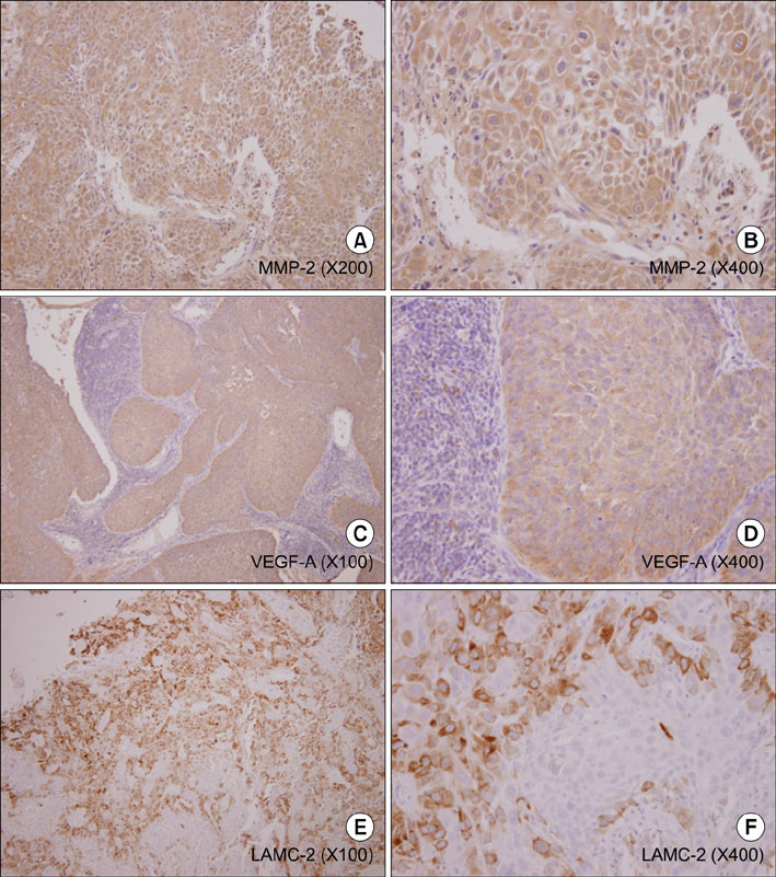
Between April 1997 and May 2008, 30 patients with disseminated cervical cancer who had supraclavicular lymph node (N=13) or hematogenous metastases (N=17) were initially treated at our institute. We reviewed medical records to extract clinicopathologic variables. For 17 patients with available pathological specimens, we evaluated the association of immunohistochemical staining for metalloproteinase (MMP)-2, vascular endothelial growth factor (VEGF)-A, and laminin V gamma (LAMC)-2 with survival and clinicopathologic variables via a log-rank test and Cox regression analysis.
RESULTS
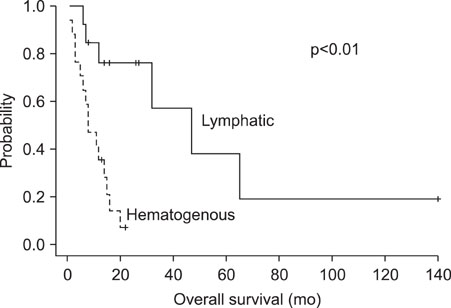
Patients who had only lymphatic metastasis (odds ratio [OR], 5.3; 95% confidence interval [CI], 1.4 to 19.5) or completed initial treatment (OR, 3.2; 95% CI, 1.1 to 9.9) showed better survival than patients who did not, but none of the molecular markers were associated with survival. Out of 13 patients with only lymphatic metastasis, three patients who had received volume-directed radiation with concurrent chemotherapy had a long-term survival of over two years. However, patients with hematogenous metastasis showed extremely poor prognosis.
CONCLUSION
The type of metastasis and completion of initial treatment were associated with prolonged survival in patients with disseminated cervical cancer, and over 20% of patients with lymphatic metastasis were salvaged with volume-directed radiation with concurrent chemotherapy. None of the molecular markers were associated with survival in patients with disseminated cervical cancer.
Keyword
MeSH Terms
Figure
Cited by 1 articles
-
Surgery of primary sites for stage IVB cervical cancer patients receiving chemoradiotherapy: a population-based study
Haoran Li, Yangyang Pang, Xi Cheng
J Gynecol Oncol. 2020;31(1):. doi: 10.3802/jgo.2020.31.e8.
Reference
-
1. IARC Cancer Epidemiology Database [Internet]. cited 2009 May 27. Lyon: International Agency for Research on Cancer;Available from: http://www-dep.iarc.fr/.2. Quinn MA, Benedet JL, Odicino F, Maisonneuve P, Beller U, Creasman WT, et al. Carcinoma of the cervix uteri. FIGO 6th Annual Report on the Results of Treatment in Gynecological Cancer. Int J Gynaecol Obstet. 2006. 95:S43–S103.3. Aziz MF. Gynecological cancer in Indonesia. J Gynecol Oncol. 2009. 20:8–10.4. Chao A, Ho KC, Wang CC, Cheng HH, Lin G, Yen TC, et al. Positron emission tomography in evaluating the feasibility of curative intent in cervical cancer patients with limited distant lymph node metastases. Gynecol Oncol. 2008. 110:172–178.5. Qiu JT, Ho KC, Lai CH, Yen TC, Huang YT, Chao A, et al. Supraclavicular lymph node metastases in cervical cancer. Eur J Gynaecol Oncol. 2007. 28:33–38.6. Van den Eynden GG, Van der Auwera I, Van Laere SJ, Trinh XB, Colpaert CG, van Dam P, et al. Comparison of molecular determinants of angiogenesis and lymphangiogenesis in lymph node metastases and in primary tumours of patients with breast cancer. J Pathol. 2007. 213:56–64.7. Van den Eynden GG, Van Laere SJ, Van der Auwera I, Gilles L, Burn JL, Colpaert C, et al. Differential expression of hypoxia and (lymph)angiogenesis-related genes at different metastatic sites in breast cancer. Clin Exp Metastasis. 2007. 24:13–23.8. Smith SC, Nicholson B, Nitz M, Frierson HF, Smolkin M, Hampton G, et al. Profiling bladder cancer organ site-specific metastasis identifies LAMC2 as a novel biomarker of hematogenous dissemination. Am J Pathol. 2009. 174:371–379.9. Kang S, Kim SK, Chung DC, Seo SS, Kim JY, Nam BH, et al. Diagnostic value of (18)F-FDG PET for evaluation of paraaortic nodal metastasis in patients with cervical carcinoma: a metaanalysis. J Nucl Med. 2010. 51:360–367.10. Horowitz NS, Tamimi HK, Goff BA, Koh WJ, Schmidt RA, Greer BE, et al. Pretreatment scalene node biopsy in gynecologic malignancy: prudent or passe? Gynecol Oncol. 1999. 75:238–241.11. Tran BN, Grigsby PW, Dehdashti F, Herzog TJ, Siegel BA. Occult supraclavicular lymph node metastasis identified by FDG-PET in patients with carcinoma of the uterine cervix. Gynecol Oncol. 2003. 90:572–576.
- Full Text Links
- Actions
-
Cited
- CITED
-
- Close
- Share
- Similar articles
-
- Angiogenesis in Cervical Intraepithelial Neoplasia and Invasive Cervical Cancer: an Immunohistochemical Study
- Spontaneous Subdural Hematoma Associated with Disseminated Intravascular Coagulation in Patient with Cancer
- Lymph Node Metastasis in a Gynecologic Malignancy
- Prognostic factors of stage IB1 cervical cancer treated surgically
- Prognostic factors in recurrent cervical cancer patients with pulmonary metastasis


