A Case of Balanced Type Double Aortic Arch Diagnosed Incidentally by Transthoracic Echocardiography in an Asymptomatic Adult Patient
- Affiliations
-
- 1Department of Internal Medicine, National Police Hospital, Seoul, Korea. bemasc@nph.go.kr
- KMID: 1980374
- DOI: http://doi.org/10.4250/jcu.2011.19.3.163
Abstract
- A 36-year-old male patient with no remarkable medical history was admitted to our hospital for a health check up. On chest radiography, bilateral aortic notches at the level of aortic arch were shown suggesting aortic arch anomaly without any clinical symptoms. Two aortic arches were almost same-in-size on suprasternal view of transthoracic echocardiography. In addition, multidetector computed tomography showed balanced type double aortic arch forming a complete vascular ring which encircled the trachea and esophagus. The trachea was slightly compressed by the vascular ring whereas the esophagus was intact. Nevertheless, the pulmonary function test was normal. The patient was discharged from hospital with instructions for periodic follow-up.
Keyword
MeSH Terms
Figure
-
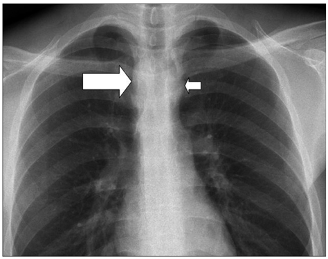
Fig. 1 Chest radiography showed bilateral aortic notches at the level of aortic arch, suggesting aortic arch anomaly. Right aortic arch (large arrow) and left aortic arch (small arrow) can be seen.

Fig. 2 Transthoracic echocardiography showed double aortic arch. Descending aorta which is located in the middle can be seen posterior to left atrium on parasternal short axis view (arrow) (A). Right aortic arch (left arrow) and left aortic arch (right arrow) (B) can be found together and respectively, right aortic arch (C), left aortic arch (D) and their branches on suprasternal view. RAA: right aortic arch, RCCA: right common carotid artery, RSCA: right subclavian artery, LAA: left aortic arch, LCCA: left common carotid artery, LSCA: left subclavian artery.
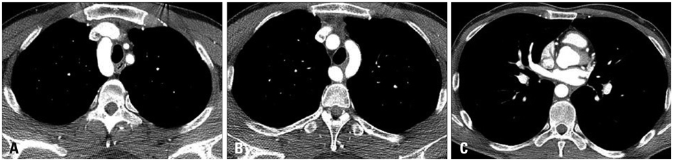
Fig. 3 Chest computed tomography showed double aortic arch. Right aortic arch (A) and left aortic arch (B) can be seen and both aortic arches fuse into descending aorta located in front of the spine (C).

Fig. 4 Reconstructed images of chest computed tomography showed balanced type double aortic arch (A) with only slight compression of the trachea (B).
Reference
-
1. Yilmaz M, Tok M, Cengiz M. Asymptomatic balanced-type double aortic arch in an elderly patient: a case report. Heart Surg Forum. 2007. 10:E297–E298.
Article2. Ito H, Konishi A, Kon-Nai T, Ishibashi T, Takahashi S. Double aortic arch with atresia, tapering and aneurysm of the left arch. Br J Radiol. 2006. 79:e71–e74.
Article3. Ikenouchi H, Tabei F, Itoh N, Nozaki A. Images in cardiovascular medicine. Silent double aortic arch found in an elderly man. Circulation. 2006. 114:e360–e361.4. Sariaydin M, Findik S, Atici AG, Ozkaya S, Uluisik A. Asymptomatic double aortic arch. Int Med Case Rep J. 2010. 3:63–66.5. Stewart JR, Kincaid OW, Edwards JE. An atlas of vascular rings and related malformations of the aortic arch system. 1963. Springfield: Thomas;579–580.6. Jeeyani HN, Prajapati VJ, Patel NH, Shah SB. Imaging features of double aortic arch shown by multidetector computed tomography angiography. Ann Pediatr Cardiol. 2010. 3:169–170.
Article7. Lowe GM, Donaldson JS, Backer CL. Vascular rings: 10-year review of imaging. Radiographics. 1991. 11:637–646.
Article8. Schlesinger AE, Krishnamurthy R, Sena LM, Guillerman RP, Chung T, DiBardino DJ, Fraser CD Jr. Incomplete double aortic arch with atresia of the distal left arch: distinctive imaging appearance. AJR Am J Roentgenol. 2005. 184:1634–1639.
Article9. Kellenberger CJ. Aortic arch malformations. Pediatr Radiol. 2010. 40:876–884.
Article10. Jaffe RB. Radiographic manifestations of congenital anomalies of the aortic arch. Radiol Clin North Am. 1991. 29:319–334.11. Moes CA, Freedom RM. Rare types of aortic arch anomalies. Pediatr Cardiol. 1993. 14:93–101.
Article12. Baraldi R, Sala S, Bighi S, Mannella P. Vascular ring due to double aortic arch: a rare cause of dysphagia. Eur J Radiol Extra. 2004. 52:21–24.
Article13. Emmel M, Schmidt B, Schickendantz S. Double aortic arch in a patient with Fallot's tetralogy. Cardiol Young. 2005. 15:52–53.
Article14. Kim HY, Jung HY, Yun TJ, Lee SS, Lee EY, Ko KH, Kim YM, Myung SJ, Yang SK, Hong WS, Kim JH, Min YI. Three cases of dysphagia due to vascular ring in adults. Korean J Gastrointest Endosc. 2000. 21:735–740.15. Lee ML. Diagnosis of the double aortic arch and its differentiation from the conotruncal malformations. Yonsei Med J. 2007. 48:818–826.
Article16. Koz C, Yokusoglu M, Uzun M, Tasar M. Double aortic arch suspected upon transthoracic echocardiography and diagnosed upon computed tomography. Tex Heart Inst J. 2008. 35:80–81.17. Lotz J, Macchiarini P. Images in clinical medicine. Double aortic arch diagnosed by magnetic resonance imaging. N Engl J Med. 2004. 351:e20.18. van Son JA, Julsrud PR, Hagler DJ, Sim EK, Puga FJ, Schaff HV, Danielson GK. Imaging strategies for vascular rings. Ann Thorac Surg. 1994. 57:604–610.
Article19. Lillehei CW, Colan S. Echocardiography in the preoperative evaluation of vascular rings. J Pediatr Surg. 1992. 27:1118–1120. discussion 1120-1.
Article20. Kypson AP, Anderson CA, Rodriguez E, Koutlas TC. Double aortic arch in an adult undergoing coronary bypass surgery: a therapeutic dilemma? Eur J Cardiothorac Surg. 2008. 34:920–921.
Article21. Alsenaidi K, Gurofsky R, Karamlou T, Williams WG, McCrindle BW. Management and outcomes of double aortic arch in 81 patients. Pediatrics. 2006. 118:e1336–e1341.
Article
- Full Text Links
-
- Actions
-
Cited
- CITED
-
- Close
- Share
-
- Similar articles
-
- A Case of Double Orifice Mitral Valve in a Patient with Bicuspid Aortic Valve: Coincidental or a Missed Finding?
- A Case of 51 Year Old Woman with Quadricuspid Aortic Valve Associated with Regurgitation
- Quadricuspid Aortic Valve: Report of Two Cases
- Quadricuspid Aortic Valve : Report of Three Cases and Review of the Literature
- Dysphagia due to Physiological Constriction after Stroke: A Case Report
