Changes in mandibular transversal arch dimensions after rapid maxillary expansion procedure assessed through cone-beam computed tomography
- Affiliations
-
- 1Department of Orthodontics, Faculty of Dentistry, Kocaeli University, Kocaeli, Turkey.
- 2Department of Orthodontics, Faculty of Dentistry, Dicle University, Diyarbakir, Turkey.
- 3Department of Orthodontics, Faculty of Dentistry, Erciyes University, Kayseri, Turkey.
- 4Department of Orthodontics, Faculty of Dentistry, Izmir Katip Celebi University, Izmir, Turkey. tancan.uysal@ikc.edu.tr
- 5Department of Pediatric Dentistry and Orthodontics, College of Dentistry, King Saud University, Riyadh, Saudi Arabia.
- KMID: 1975432
- DOI: http://doi.org/10.4041/kjod.2011.41.3.200
Abstract
OBJECTIVE
This study aimed at evaluating the changes in mandibular arch widths and buccolingual inclinations of mandibular posterior teeth after rapid maxillary expansion (RME).
METHODS
Baseline and post-expansion cone-beam computed tomographic (CBCT) images of patients who initially had bilateral posterior cross-bite and underwent RME with a banded-type expander were assessed in this study. The patients included 9 boys (mean age: 13.97 +/- 1.17 years) and 11 girls (mean age: 13.53 +/- 2.12 years). Images obtained 6 months after retention were available for 10 of these patients. Eighteen angular and 43 linear measurements were performed for the maxilla and mandible. The measurements were performed on frontally clipped images at the following time points; before expansion (T1), after expansion (T2), and after retention (T3). Statistical significance was assessed with paired sample t-test at p < 0.05.
RESULTS
T1-T2 comparisons showed statistically significant post-RME increases for all measurements; similarly, T2-T1 and T3-T1 comparisons showed statistically significant changes. The maxillary linear and angular measurements showed decreases after expansion, and mandibular linear and angular measurements increased after retention.
CONCLUSION
All mandibular arch widths increased and mandibular posterior teeth were uprighted after RME procedure.
MeSH Terms
Figure
-
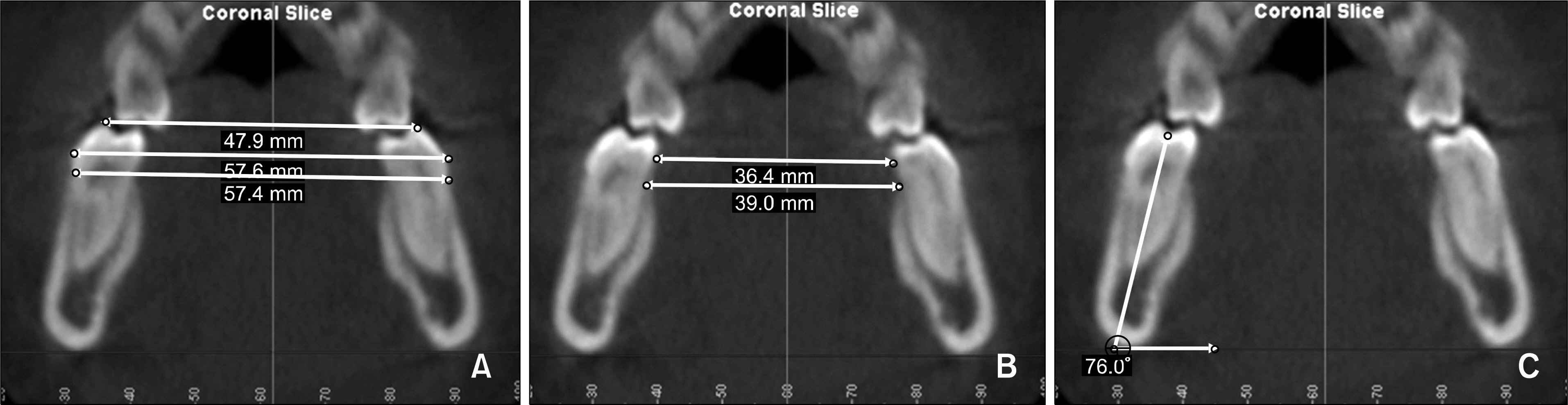
Fig 1. Mandibular linear and angular measurements. A, External mandibular measurements performed at 3 different levels: alveolar crest, most prominent point of the crown, and buccal cusp tip; B, internal mandibular measurements were performed at 2 different levels: alveolar crest and most prominent point of the crown; C, mandibular long axis measurement performed between the long axis of the mesial root and to the reference plane, which passes through the most prominent point of the lower borders (right and left) of the mandible.
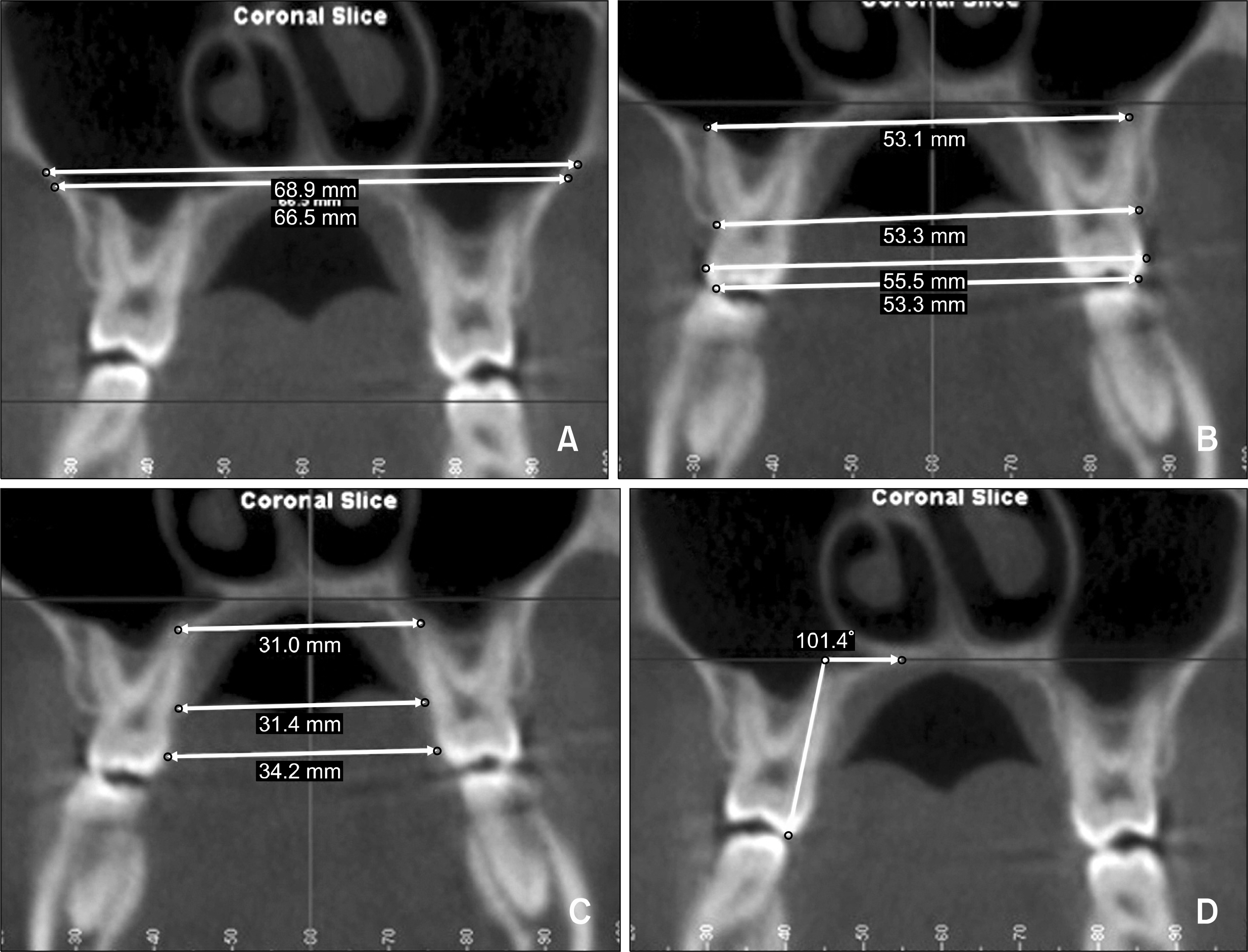
Fig 2. Maxillary linear and angular measurements. A, Nasal and palatal floor measurements; B, external maxillary measurements performed at 4 different levels: apical, alveolar crest, most prominent point of the crown, and buccal cusp tip; C, internal maxillary measurements were performed at 3 different levels: apical, alveolar crest, and most prominent point of the crown; D, maxillary long axis measurement performed between the long axis of the palatal root and to the palatal plane.
Cited by 1 articles
-
Short-term treatment effects produced by rapid maxillary expansion evaluated with computed tomography: A systematic review with meta-analysis
Antonino Lo Giudice, Paola Spinuzza, Lorenzo Rustico, Gabriele Messina, Riccardo Nucera
Korean J Orthod. 2020;50(5):314-323. doi: 10.4041/kjod.2020.50.5.314.
Reference
-
1.Angell EC. Treatment of irregularity of the permanent or adult teeth. Dent Cosmos. 1860. 1:540–4.2.Miller CL. Concomitant changes in mandibular arch dimensions during bonded and banded rapid maxillary expansion (dissertation). Missouri: Saint Louis University. 2010.3.Haas AJ. Rapid expansion of the maxillary dental arch and nasal cavity by opening the mid-palatal suture. Angle Orthod. 1961. 31:73–90.4.Haas AJ. The treatment of maxillary deficiency by opening the midpalatal suture. Angle Orthod. 1965. 35:200–17.5.Haas AJ. Palatal expansion: just the beginning of dentofacial orthopedics. Am J Orthod. 1970. 57:219–55.
Article6.Wertz RA. Skeletal and dental changes accompanying rapid midpalatal suture opening. Am J Orthod. 1970. 58:41–66.
Article7.Bishara SE., Staley RN. Maxillary expansion: clinical implications. Am J Orthod Dentofacial Orthop. 1987. 91:3–14.8.Byrum AG Jr. Evaluation of anterior-posterior and vertical skeletal change vs. dental change in rapid palatal expansion cases as studied by lateral cephalograms. Am J Orthod. 1971. 60:419.
Article9.Hicks EP. Slow maxillary expansion. A clinical study of the skeletal versus dental response to low magnitude force. Am J Orthod. 1978. 73:121–41.10.Brodie AG. The fourth dimension in orthodontia. Angle Orthod. 1954. 24:15–30.11.Gryson JA. Changes in mandibular interdental distance concurrent with rapid maxillary expansion. Angle Orthod. 1977. 47:186–92.12.Lima AC., Lima AL., Filho RM., Oyen OJ. Spontaneous mandibular arch response after rapid palatal expansion: a long-term study on Class I malocclusion. Am J Orthod Dentofacial Orthop. 2004. 126:576–82.
Article13.Scarfe WC., Farman AG., Sukovic P. Clinical applications of cone-beam computed tomography in dental practice. J Can Dent Assoc. 2006. 72:75–80.14.Kartalian A., Gohl E., Adamian M., Enciso R. Cone-beam computerized tomography evaluation of the maxillary dentoskeletal complex after rapid palatal expansion. Am J Orthod Dentofacial Orthop. 2010. 138:486–92.
Article15.Baker LW. The influence of the formative dental organs on the growth of the bones of the face. Am J Orthod. 1941. 27:489–506.
Article16.Isaacson RJ., Wood JL., Ingram AH. Forces produced by rapid maxillary expansion. Angle Orthod. 1964. 34:256–70.17.Sandstrom RA., Klapper L., Papaconstantinou S. Expansion of the lower arch concurrent with rapid maxillary expansion. Am J Orthod Dentofacial Orthop. 1988. 94:296–302.
Article18.Geran RG., McNamara JA Jr., Baccetti T., Franchi L., Shapiro LM. A prospective long-term study on the effects of rapid maxillary expansion in the early mixed dentition. Am J Orthod Dentofacial Orthop. 2006. 129:631–40.
Article19.Moyers RE., van der Linden FPGM., Riolo ML., McNamara JA Jr. Standards of Human Occlusal Development. Monograph 5. Craniofacial Growth Series. Ann Arbor: Center for Human Growth and Development, University of Michigan;1976. p. 49–157.20.Moorrees CF., Reed RB. Changes in dental arch dimensions expressed on the basis of tooth eruption as a measure of biologic age. J Dent Res. 1965. 44:129–41.
Article21.Haas AJ. Long-term posttreatment evaluation of rapid palatal expansion. Angle Orthod. 1980. 50:189–217.22.Moussa R., O'Reilly MT., Close JM. Long-term stability of rapid palatal expander treatment and edgewise mechanotherapy. Am J Orthod Dentofacial Orthop. 1995. 108:478–88.
Article23.Glenn G., Sinclair PM., Alexander RG. Nonextraction orthodontic therapy: posttreatment dental and skeletal stability. Am J Orthod Dentofacial Orthop. 1987. 92:321–8.
Article24.Lagravère MO., Heo G., Major PW., Flores-Mir C. Meta-analysis of immediate changes with rapid maxillary expansion treatment. J Am Dent Assoc. 2006. 137:44–53.
Article
- Full Text Links
-
- Actions
-
Cited
- CITED
-
- Close
- Share
-
- Similar articles
-
- Three-dimensional assessment of the temporomandibular joint and mandibular dimensions after early correction of the maxillary arch form in patients with Class II division 1 or division 2 malocclusion
- Evaluation of condylar dimension and position following rapid maxillary expansion with toothor tooth-bone-borne appliances
- Maxillary sinus pneumatization after maxillary molar extraction assessed with cone beam computed tomography
- A cone-beam computed tomography evaluation of buccal bone thickness following maxillary expansion
- Expansion of the mandibular arch using a trombone appliance
