Finite element analysis of effectiveness of lever arm in lingual sliding mechanics
- Affiliations
-
- 1Private Practice, Korea.
- 2Department of Orthodontics, College of Dentistry, Yonsei University, Korea. ypark@yuhs.ac
- KMID: 1975387
- DOI: http://doi.org/10.4041/kjod.2011.41.5.324
Abstract
OBJECTIVE
The aim of this study was to conduct three-dimensional finite element analysis of individual tooth displacement and stress distribution when a posterior retraction force of 200 g was applied at different positions of the retraction hook on the transpalatal arch (TPA) of a molar, and over different lengths of the lever arm on the maxillary anterior teeth in lingual orthodontics.
METHODS
A three-dimensional finite element model, including the entire upper dentition, periodontal ligaments, and alveolar bones, was constructed on the basis of a sample (Nissan Dental Product, Kyoto, Japan) survey of Asian adults. Individual movement of the incisal edge and root apex was estimated along the x-, y-, and z-coordinates to analyze tooth displacement and von Mises stress distribution.
RESULTS
When the length of the lever arm was 15 mm and 20 mm, the incisal edge and root apex of the anterior teeth was displaced lingually, with a maximum lingual displacement at the lever arm length of 20 mm. When the posterior retraction hook was on the root apex, the molars showed distal displacement. When the length of the lever arm was 20 mm, anterior extrusion was reduced and the crown of the canine displaced toward the buccal side, in which case, the retraction hook was on the edge, rather than at the center, of the TPA.
CONCLUSIONS
The results of the analysis showed that when 6 anterior teeth were retracted posteriorly, lateral displacement of the canine and lingual displacement of the incisal edge and root apex of the anterior teeth occur without the extrusion of the anterior segment when the length of the lever arm is longer, and the posterior retraction hook is in the midpalatal area.
MeSH Terms
Figure
-
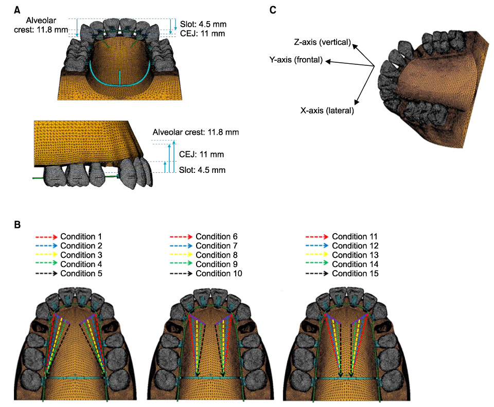
Fig. 1 A, Three-dimensional finite element model for upper teeth, periodontal ligaments and alveolar bones; B, experimental groups are divided into three groups depending on the position of retraction: position of retraction arm in the first group is placed at the bracket of the 2nd molar, at the furcation of the 2nd molar in the second group and at the root apex of the 2nd molar in the third group; C, the coordinate system is composed of X-axis, Y-axis and Z-axis. The origin of coordinate axes is the middle point of the incisal surface which connects with the upper right and left incisors. X-axis is the bucco-palatal direction (lingual (+), buccal (-)); Y-axis is the antero-posterior direction (anterior (+), posterior (-)); Z-axis is the superior-inferior direction (superior (+), inferior direction (-)). CEJ, Cemento enamel junction.
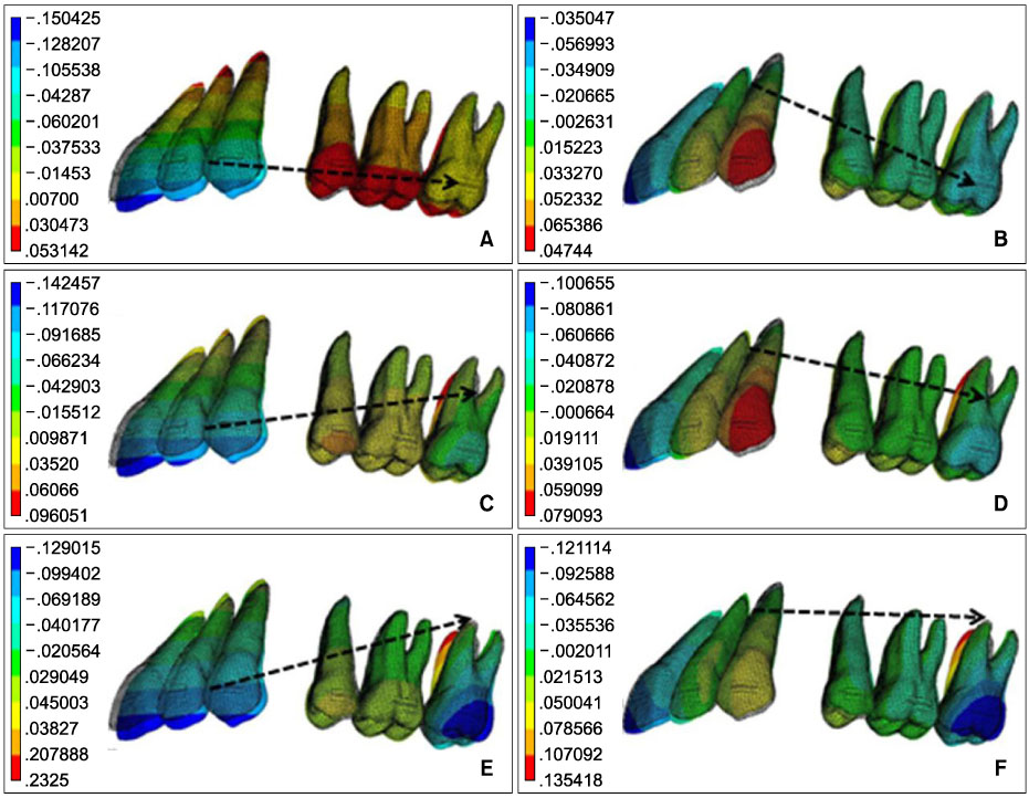
Fig. 2 Contour plot of frontal displacement at each condition. A, Length of lever arm is 0 mm and position of retraction hook is at the bracket of the 2nd molar; B, length of lever arm is 20 mm and position of retraction hook is at the bracket of the 2nd molar; C, length of lever arm is 0 mm and position of retraction hook is at the furcation of the 2nd molar; D, length of lever arm is 20 mm and position of retraction hook is at the furcation of the 2nd molar; E, length of lever arm is 0 mm and position of retraction hook is at the root apex of the 2nd molar; F, length of lever arm is 20 mm and position of retraction hook is at the root apex of the 2nd molar.

Fig. 3 Contour plot of vertical displacement at each condition. A, Length of lever arm is 0 mm and position of retraction hook is at the bracket of the 2nd molar; B, length of lever arm is 20 mm and position of retraction hook is at the bracket of the 2nd molar; C, length of lever arm is 0 mm and position of retraction hook is at the furcation of the 2nd molar; D, length of lever arm is 20 mm and position of retraction hook is at the furcation of the 2nd molar; E, length of lever arm is 0 mm and position of retraction hook is at the root apex of the 2nd molar; F, length of lever arm is 20 mm and position of retraction hook is at the root apex of the 2nd molar.

Fig. 4 Contour plot of lateral displacement at each condition. A, Length of lever arm is 0 mm and position of retraction hook is at the bracket of the 2nd molar; B, length of lever arm is 20 mm and position of retraction hook is at the bracket of the 2nd molar; C, length of lever arm is 0 mm and position of retraction hook is at the furcation of the 2nd molar; D, length of lever arm is 20 mm and position of retraction hook is at the furcation of the 2nd molar; E, length of lever arm is 0 mm and position of retraction hook is at the root apex of the 2nd molar; F, length of lever arm is 20 mm and position of retraction hook is at the root apex of the 2nd molar.
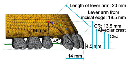
Fig. 5 Comparison of the position of lever arm and CR of anterior teeth from other studies35: (blue) - study by Jeong et al35 showing center of resistance of six anterior teeth; (green) - study by Vanden Bulcke et al31 showing center of resistance of four anterior teeth; (purple) - study by Vanden Bulcke et al31 showing center of resistance of six anterior teeth. CR, Center of resistance; CEJ, cemento enamel junction.
Cited by 2 articles
-
Torque control during lingual anterior retraction without posterior appliances
Sung-Seo Mo, Seong-Hun Kim, Sang-Jin Sung, Kyu-Rhim Chung, Yun-Sic Chun, Yoon-Ah Kook, Gerald Nelson
Korean J Orthod. 2013;43(1):3-14. doi: 10.4041/kjod.2013.43.1.3.Comparison of inclination and vertical changes between single-wire and double-wire retraction techniques in lingual orthodontics
Bui Quang Hung, Mihee Hong, Wonjae Yu, Hee-Moon Kyung
Korean J Orthod. 2020;50(1):26-32. doi: 10.4041/kjod.2020.50.1.26.
Reference
-
1. Liang W, Rong Q, Lin J, Xu B. Torque control of the maxillary incisors in lingual and labial orthodontics: a 3-dimensional finite element analysis. Am J Orthod Dentofacial Orthop. 2009; 135:316–322.
Article2. Kucher G, Weiland FJ, Bantleon HP. Modified lingual lever arm technique. J Clin Orthod. 1993; 27:18–22.3. Park YC, Choy K, Lee JS, Kim TK. Lever-arm mechanics in lingual orthodontics. J Clin Orthod. 2000; 34:601–605.4. Sia S, Koga Y, Yoshida N. Determining the center of resistance of maxillary anterior teeth subjected to retraction forces in sliding mechanics. An in vivo study. Angle Orthod. 2007; 77:999–1003.
Article5. Lewis G, Kambhampati S, Roussel S. Effect of the archwire slot profile on the performance of bonded orthodontic brackets. Biomed Mater Eng. 1997; 7:205–212.
Article6. Burstone CJ, Pryputniewicz RJ. Holographic determination of centers of rotation produced by orthodontic forces. Am J Orthod. 1980; 77:396–409.
Article7. Pedersen E, Andersen K, Melsen B. Tooth displacement analysed on human autopsy material by means of a strain gauge technique. Eur J Orthod. 1991; 13:65–74.
Article8. Reimann S, Keilig L, Jäger A, Bourauel C. Biomechanical finite-element investigation of the position of the centre of resistance of the upper incisors. Eur J Orthod. 2007; 29:219–224.
Article9. Kim CN, Sung JH, Kyung HM. Three-dimensional finite element analysis of initial tooth displacement according to force application point during maxillary six anterior teeth retraction using skeletal anchorage. Korean J Orthod. 2003; 33:339–350.10. Andrews LF. The six keys to normal occlusion. Am J Orthod. 1972; 62:296–309.
Article11. Coolidge E. The thickness of the human periodontal membrane. J Am Dent Assoc. 1937; 24:1260–1265.
Article12. Kronfeld R. Histologic study of the influence of function on the human periodontal membrane. J Am Dent Assoc. 1931; 18:1242–1272.13. Andrews LF. Andrews LF, editor. The six keys to optimal occlusion. Straight wire: the concept and appliance. 1989. San Diego: LA Wells;p. 13–24.14. Cook SD, Weinstein AM, Klawitter JJ. A three-dimensional finite element analysis of a porous rooted Co-Cr-Mo alloy dental implant. J Dent Res. 1982; 61:25–29.15. Tanne K, Sakuda M, Burstone CJ. Three-dimensional finite element analysis for stress in the periodontal tissue by orthodontic forces. Am J Orthod Dentofacial Orthop. 1987; 92:499–505.
Article16. Sung SJ, Baik HS, Moon YS, Yu HS, Cho YS. A comparative evaluation of different compensating curves in the lingual and labial techniques using 3D FEM. Am J Orthod Dentofacial Orthop. 2003; 123:441–450.
Article17. Bennett JC, McLaughlin RP. Controlled space closure with a preadjusted appliance system. J Clin Orthod. 1990; 24:251–260.18. Alexander CM, Alexander RG, Gorman JC, Hilgers JJ, Kurz C, Scholz RP, et al. Lingual orthodontics: a status report. Part 5. Lingual mechanotherapy. J Clin Orthod. 1983; 17:99–115.19. Smith JR, Gorman JC, Kurz C, Dunn RM. Keys to success in lingual therapy. Part 1. J Clin Orthod. 1986; 20:252–261.20. Smith RJ, Burstone CJ. Mechanics of tooth movement. Am J Orthod. 1984; 85:294–307.
Article21. Davidian EJ. Use of a computer model to study the force distribution on the root of the maxillary central incisor. Am J Orthod. 1971; 59:581–588.
Article22. Burstone CJ, Koenig HA. Optimizing anterior and canine retraction. Am J Orthod. 1976; 70:1–19.
Article23. Vanden Bulcke MM, Burstone CJ, Sachdeva RC, Dermaut LR. Location of the centers of resistance for anterior teeth during retraction using the laser reflection technique. Am J Orthod Dentofacial Orthop. 1987; 91:375–384.
Article24. Woo JY, Park YC. Experimental study of the vertical location of the centers of resistance for maxillary anterior teeth during retraction using the laser reflection technique. Korean J Orthod. 1993; 23:375–389.25. Park GH, Sohn BW. The center of resistance of the maxillary anterior segment in the horizontal plane during intrusion by using laser reflection technique. Korean J Orthod. 1993; 23:619–631.26. Lee HK, Chung KR. The vertical location of the center of resistance for maxillary six anterior teeth during retraction using three dimensional finite element analysis. Korean J Orthod. 2001; 31:425–438.27. Jeong GM, Sung SJ, Lee KJ, Chun YS, Mo SS. Finite-element investigation of the center of resistance of the maxillary dentition. Korean J Orthod. 2009; 39:83–94.
Article28. Tanne K, Yoshida S, Kawata T, Sasaki A, Knox J, Jones ML. An evaluation of the biomechanical response of the tooth and periodontium to orthodontic forces in adolescent and adult subjects. Br J Orthod. 1998; 25:109–115.
Article29. Andersen KL, Pedersen EH, Melsen B. Material parameters and stress profiles within the periodontal ligament. Am J Orthod Dentofacial Orthop. 1991; 99:427–440.
Article30. Cobo J, Argüelles J, Puente M, Vijande M. Dentoalveolar stress from bodily tooth movement at different levels of bone loss. Am J Orthod Dentofacial Orthop. 1996; 110:256–262.
Article
- Full Text Links
-
- Actions
-
Cited
- CITED
-
- Close
- Share
-
- Similar articles
-
- Finite element analysis of the effects of different archwire forms and power arm positions on maxillary incisors in en masse retraction using fixed lingual orthodontic appliances
- Three dimensional finite element analysis of continuous and segmented arches with use of orthodontic miniscrews
- Palatal en-masse retraction of segmented maxillary anterior teeth: A finite element study
- A photoelastic study on the initial stress distribution of the upper anterior teeth retraction using combination loop archwire and sliding mechanics
- Effect of archwire stiffness and friction on maxillary posterior segment displacement during anterior segment retraction: A three-dimensional finite element analysis
