Korean J Orthod.
2024 Sep;54(5):265-273. 10.4041/kjod23.196.
Finite element analysis of the effects of different archwire forms and power arm positions on maxillary incisors in en masse retraction using fixed lingual orthodontic appliances
- Affiliations
-
- 1Department of Orthodontics, Usak University, Usak, Türkiye
- 2Private Practice, Izmir, Türkiye
- KMID: 2559816
- DOI: http://doi.org/10.4041/kjod23.196
Abstract
Objective
This study aimed to investigate the effects of archwire form and power arm positions on maxillary incisors during lingual en masse retraction supported by miniscrew implants, using the finite element analysis method.
Methods
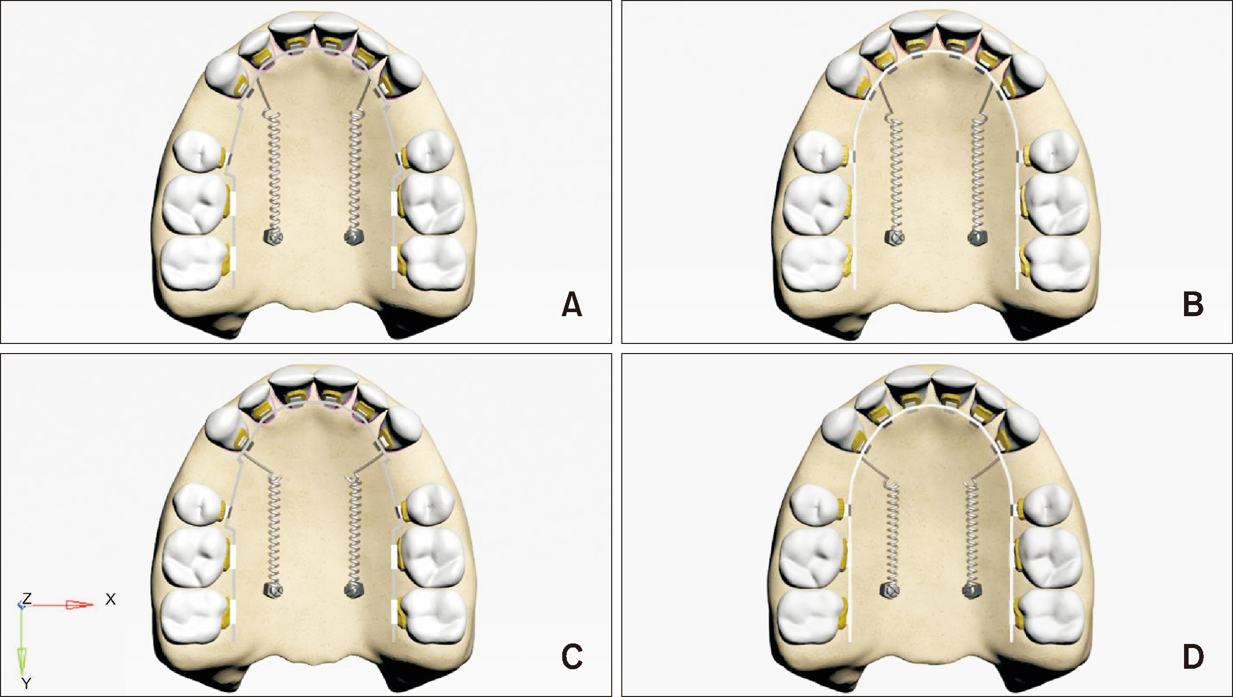
Sliding mechanics for lingual en masse retraction were simulated using the finite element method. Power arms were placed mesial and distal to the maxillary canine with straight and mushroom-shaped archwires. Miniscrews provided absolute anchorage for retraction force.
Results
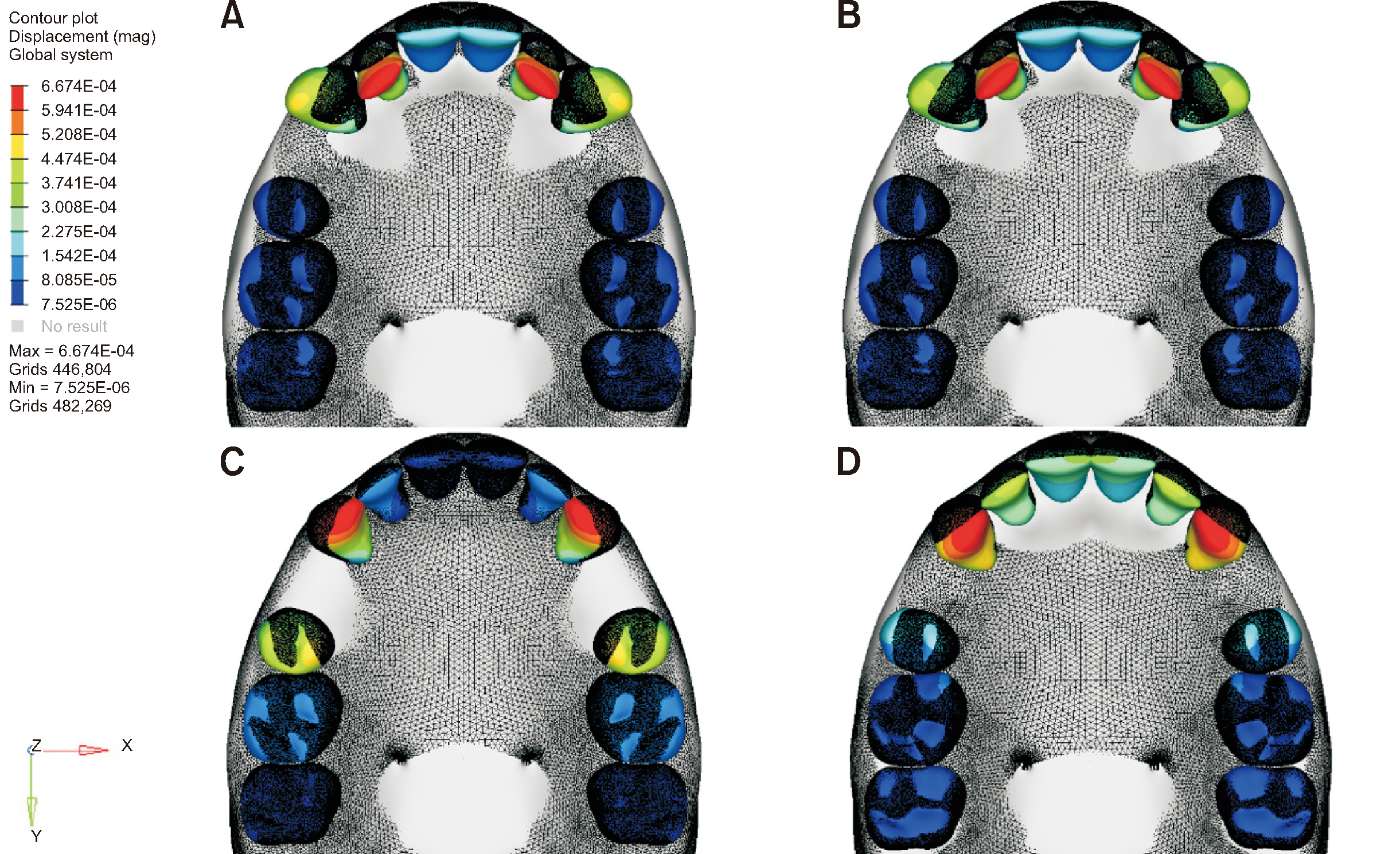
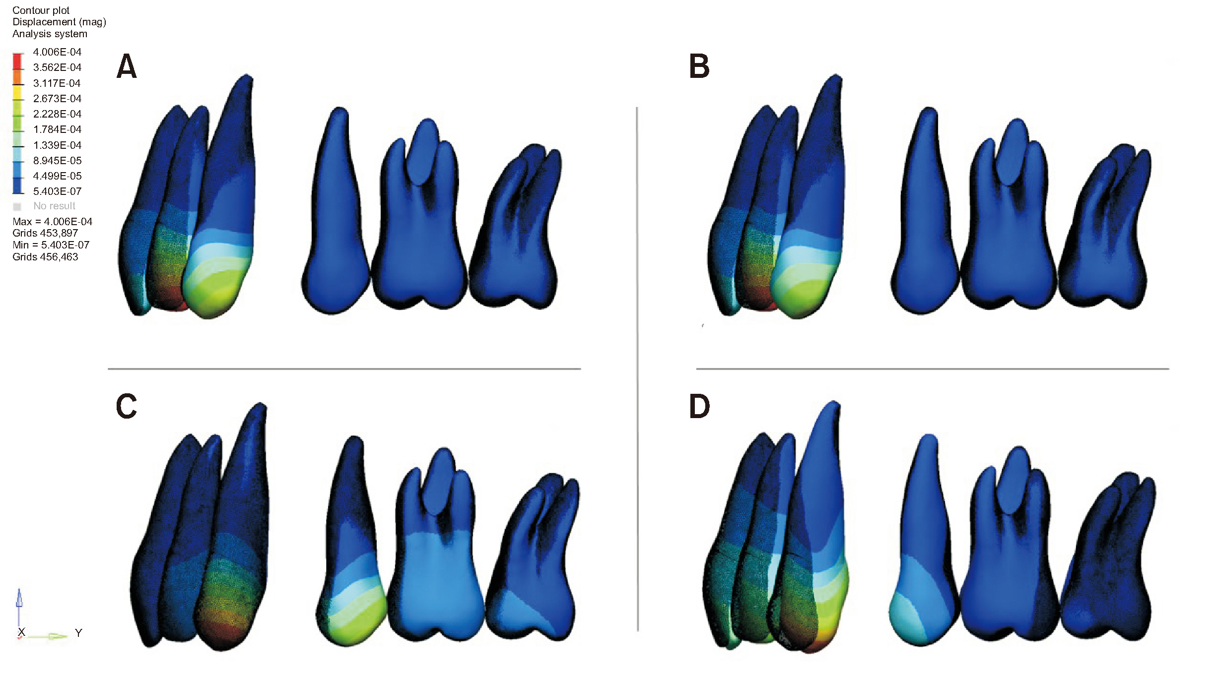
When power arms were positioned mesial to the canine teeth, an increase in the intercanine distance was observed, while a decrease was noted when the power arms were distal to the canine tooth. Lateral incisors exhibited a greater torque loss, particularly when the power arm was mesial to the canine tooth. In the central incisors, the mushroom archwire resulted in intrusion, while the straight archwire showed an extrusion tendency. Movements in groups using the straight archwire were less controlled compared to those in groups using the mushroom archwire.
Conclusions
The archwire form and the position of the power arm affected the torque loss and vertical position of incisors during lingual en masse retraction supported by miniscrew implants. The most controlled movement was achieved with the combination of a power arm positioned distal to the canine tooth and a mushroom archform.
Figure
Reference
-
1. Fujita K. 1979; New orthodontic treatment with lingual bracket mushroom arch wire appliance. Am J Orthod. 76:657–75. https://doi.org/10.1016/0002-9416(79)90211-2. DOI: 10.1016/0002-9416(79)90211-2.2. Scuzzo G, Takemoto K, Takemoto Y, Takemoto A, Lombardo L. 2010; A new lingual straight-wire technique. J Clin Orthod. 44:114–23. quiz 106https://pubmed.ncbi.nlm.nih.gov/20552813/.3. Nanda R, Kuhlberg A. Nanda R, editor. 1997. Principles of biomechanics. Biomechanics in clinical orthodontics. Saunders Book Company;Collingwood: p. 188–217. https://www.amazon.com/Biomechanics-Clinical-Orthodontics-Ravindra-Nanda/dp/0721627846.4. Moran KI. 1987; Relative wire stiffness due to lingual versus labial interbracket distance. Am J Orthod Dentofacial Orthop. 92:24–32. https://doi.org/10.1016/0889-5406(87)90292-7. DOI: 10.1016/0889-5406(87)90292-7.5. Thurow RC. 1975; Letter: elastic ligatures, binding forces, and anchorage taxation. Am J Orthod. 67:694. https://doi.org/10.1016/0002-9416(75)90146-3. DOI: 10.1016/0002-9416(75)90146-3. PMID: 1056140.6. Andreasen GF, Quevedo FR. 1970; Evaluation of friction forces in the 0.022 x 0.028 edgewise bracket in vitro. J Biomech. 3:151–60. https://doi.org/10.1016/0021-9290(70)90002-3. DOI: 10.1016/0021-9290(70)90002-3.7. Kushwah A, Kumar M, Goyal M, Premsagar S, Rani S, Sharma S. 2020; Analysis of stress distribution in lingual orthodontics system for effective en-masse retraction using various combinations of lever arm and mini-implants: a finite element method study. Am J Orthod Dentofacial Orthop. 158:e161–72. https://doi.org/10.1016/j.ajodo.2020.08.005. DOI: 10.1016/j.ajodo.2020.08.005. PMID: 33250107.8. Khosravi R. 2018; Biomechanics in lingual orthodontics: what the future holds. Semin Orthod. 24:363–71. https://doi.org/10.1053/j.sodo.2018.08.008. DOI: 10.1053/j.sodo.2018.08.008.9. Hong RK, Heo JM, Ha YK. 2005; Lever-arm and mini-implant system for anterior torque control during retraction in lingual orthodontic treatment. Angle Orthod. 75:129–41. https://pubmed.ncbi.nlm.nih.gov/15747828/.10. Liang W, Rong Q, Lin J, Xu B. 2009; Torque control of the maxillary incisors in lingual and labial orthodontics: a 3-dimensional finite element analysis. Am J Orthod Dentofacial Orthop. 135:316–22. https://doi.org/10.1016/j.ajodo.2007.03.039. DOI: 10.1016/j.ajodo.2007.03.039.11. Scuzzo G, Takemoto K. Scuzzo G, Takemoto K, editors. 2003. Biomechanics and comparative biomechanics. Invisible orthodontics. Quintessence Books;Berlin: p. 55–9. https://books.google.co.kr/books/about/Invisible_Orthodontics.html?id=VQZqAAAAMAAJ&redir_esc=y. DOI: 10.1007/978-3-031-49204-4.12. Matsui S, Caputo AA, Chaconas SJ, Kiyomura H. 2000; Center of resistance of anterior arch segment. Am J Orthod Dentofacial Orthop. 118:171–8. https://doi.org/10.1067/mod.2000.103774. DOI: 10.1067/mod.2000.103774.13. Choy K, Kim KH, Burstone CJ. 2006; Initial changes of centres of rotation of the anterior segment in response to horizontal forces. Eur J Orthod. 28:471–4. https://doi.org/10.1093/ejo/cjl023. DOI: 10.1093/ejo/cjl023.14. Kim KY, Bayome M, Park JH, Kim KB, Mo SS, Kook YA. 2015; Displacement and stress distribution of the maxillofacial complex during maxillary protraction with buccal versus palatal plates: finite element analysis. Eur J Orthod. 37:275–83. https://doi.org/10.1093/ejo/cju039. DOI: 10.1093/ejo/cju039.15. Feng Y, Kong WD, Cen WJ, Zhou XZ, Zhang W, Li QT, et al. 2019; Finite element analysis of the effect of power arm locations on tooth movement in extraction space closure with miniscrew anchorage in customized lingual orthodontic treatment. Am J Orthod Dentofacial Orthop. 156:210–9. https://doi.org/10.1016/j.ajodo.2018.08.025. DOI: 10.1016/j.ajodo.2018.08.025.16. Cattaneo PM, Dalstra M, Melsen B. 2005; The finite element method: a tool to study orthodontic tooth movement. J Dent Res. 84:428–33. https://doi.org/10.1177/154405910508400506. DOI: 10.1177/154405910508400506.17. Chung KR, Kim SH, Kook YA, Son JH. 2008; Anterior torque control using partial-osseointegrated mini-implants: biocreative therapy type I technique. World J Orthod. 9:95–104. https://pubmed.ncbi.nlm.nih.gov/18575303/.18. Chung KR, Kim SH, Kook YA, Choo H. 2008; Anterior torque control using partial-osseointegrated mini-implants: biocreative therapy type II technique. World J Orthod. 9:105–13. https://pubmed.ncbi.nlm.nih.gov/18575304/.19. Kim JS, Kim SH, Kook YA, Chung KR, Nelson G. 2011; Analysis of lingual en masse retraction combining a C-lingual retractor and a palatal plate. Angle Orthod. 81:662–9. https://doi.org/10.2319/100110-574.1. DOI: 10.2319/100110-574.1.20. Mo SS, Kim SH, Sung SJ, Chung KR, Chun YS, Kook YA, et al. 2013; Torque control during lingual anterior retraction without posterior appliances. Korean J Orthod. 43:3–14. https://doi.org/10.4041/kjod.2013.43.1.3. DOI: 10.4041/kjod.2013.43.1.3. PMID: 23502971. PMCID: PMC3594878.21. Chung KR, Kook YA, Kim SH, Mo SS, Jung JA. 2008; Class II malocclusion treated by combining a lingual retractor and a palatal plate. Am J Orthod Dentofacial Orthop. 133:112–23. https://doi.org/10.1016/j.ajodo.2006.04.033. DOI: 10.1016/j.ajodo.2006.04.033.22. Basha AG, Shantaraj R, Mogegowda SB. 2010; Comparative study between conventional en-masse retraction (sliding mechanics) and en-masse retraction using orthodontic micro implant. Implant Dent. 19:128–36. https://doi.org/10.1097/ID.0b013e3181cc4aa5. DOI: 10.1097/ID.0b013e3181cc4aa5.23. Sung SJ, Jang GW, Chun YS, Moon YS. 2010; Effective en-masse retraction design with orthodontic mini-implant anchorage: a finite element analysis. Am J Orthod Dentofacial Orthop. 137:648–57. https://doi.org/10.1016/j.ajodo.2008.06.036. DOI: 10.1016/j.ajodo.2008.06.036. PMID: 20451784.24. Mattu N, Virupaksha AM, Belludi A. 2021; Comparative study of effect of different lever arm positions and lengths on transverse and vertical bowing in lingual orthodontics - an FEM study. Int Orthod. 19:281–90. https://doi.org/10.1016/j.ortho.2021.03.003. Erratum in: Int Orthod 2024;22:100850. https://doi.org/10.1016/j.ortho.2024.100850. DOI: 10.1016/j.ortho.2024.100850.25. Sadek MM, Sabet NE, Hassan IT. 2019; Type of tooth movement during en masse retraction of the maxillary anterior teeth using labial versus lingual biocreative therapy in adults: a randomized clinical trial. Korean J Orthod. 49:381–92. https://doi.org/10.4041/kjod.2019.49.6.381. DOI: 10.4041/kjod.2019.49.6.381.26. Hung BQ, Hong M, Yu W, Kyung HM. 2020; Comparison of inclination and vertical changes between single-wire and double-wire retraction techniques in lingual orthodontics. Korean J Orthod. 50:26–32. https://doi.org/10.4041/kjod.2020.50.1.26. DOI: 10.4041/kjod.2020.50.1.26. PMID: 32042717. PMCID: PMC6995831.27. Hedayati Z, Shomali M. 2016; Maxillary anterior en masse retraction using different antero-posterior position of mini screw: a 3D finite element study. Prog Orthod. 17:31. https://doi.org/10.1186/s40510-016-0143-z. DOI: 10.1186/s40510-016-0143-z. PMID: 27667816. PMCID: PMC5045917. PMID: 3049a83e2aff4947bae7a919c71ec757.28. Zalaquett R, Karam R, Kaddah F, Khoury E, El Khoury T, Ghoubril J, et al. 2024; Effect of power arm length combined with additional anterior torque on the axial orientation of the maxillary incisors during en-masse retraction: a finite element analysis. Am J Orthod Dentofacial Orthop. 165:220–31. https://doi.org/10.1016/j.ajodo.2023.08.016. DOI: 10.1016/j.ajodo.2023.08.016.
- Full Text Links
- Actions
-
Cited
- CITED
-
- Close
- Share
- Similar articles
-
- Factors influencing the axes of anterior teeth during SWA en masse sliding retraction with orthodontic mini-implant anchorage: a finite element study
- Three-dimensional finite element analysis of the phenomenon produced during retraction of four maxillary incisors
- Three dimensional finite element analysis of the phenomenon during distal en masse movement of the maxillary dentition
- Force changes associated with differential activation of en-masse retraction and/or intrusion with clear aligners
- Palatal en-masse retraction of segmented maxillary anterior teeth: A finite element study


