J Adv Prosthodont.
2011 Jun;3(2):96-100. 10.4047/jap.2011.3.2.96.
Full mouth implant rehabilitation of a patient with ectodermal dysplasia after orthognathic surgery, sinus and ridge augmentation: a clinical report
- Affiliations
-
- 1Craniomaxillofacial Research Center, Shariati Hospital, Tehran University of Medical Sciences, Tehran, Iran. f_m_heravi@yahoo.com
- 2School of Dentistry, Shahid Beheshti University, Tehran, Iran.
- 3School of Dentistry, Hamedan University of Medical Sciences, Iran.
- KMID: 1975084
- DOI: http://doi.org/10.4047/jap.2011.3.2.96
Abstract
- An 18-year-old male presented severe hypodontia due to hypohidrotic ectodermal dysplasia was treated with Le Fort I maxillary osteotomy with simultaneous sinus floor augmentation using the mixture of cortical autogenous bone graft harvested from iliac crest and organic Bio-Oss to position the maxilla in a right occlusal plane with respect to the mandible, and to construct adequate bone volume at posterior maxilla allowing proper implant placement. Due to the poor bone quality at other sites, ridge augmentation with onlay graft was done to construct adequate bone volume allowing proper implant placement, using tissue harvested from the iliac bone. Seven implants were placed in the maxilla and 7 implants were inserted in the mandible and screw-retained metal ceramic FPDs were fabricated. The two year follow up data showed that dental implants should be considered as a good treatment modality for patients with ectodermal dysplasia.
MeSH Terms
Figure
-

Fig. 1 A, B: Profile of the patient preoperatively.

Fig. 2 Pretreatment intraoral view showing disproportionate vertical dimension of occlusion, and underdeveloped alveolar ridges for implant rehabilitation.
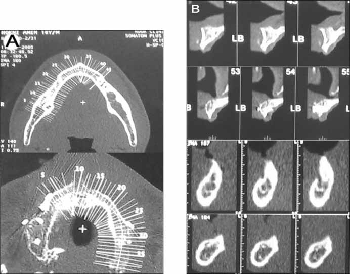
Fig. 3 A, B: Pretreatment CT scan finding of the study prior to the bone augmentation.
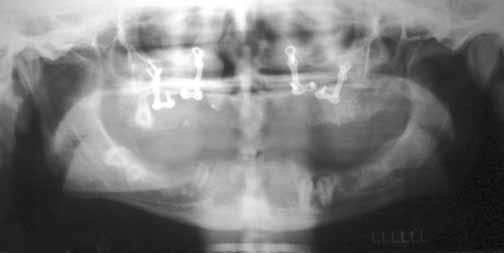
Fig. 4 Panoramic view of patient after orthognathic surgery.
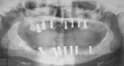
Fig. 5 Panoramic view after implant insertion and graft surgery showing positions of maxillary and mandibular dental implants.
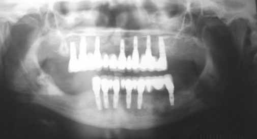
Fig. 6 Panoramic view taken 24 months after delivery of final restoration.
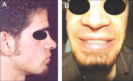
Fig. 7 A, B: Patient's profiles.
Reference
-
1. Bergendal B. Oligodontia ectodermal dysplasia-on signs, symptoms, genetics, and outcomes of dental treatment. Swed Dent J Suppl. 2010. 205:13–78. 7–8.2. Van Sickels JE, Raybould TP, Hicks EP. Interdisciplinary management of patients with ectodermal dysplasia. J Oral Implantol. 2010. 36:239–245.3. Sclar AG, Kannikal J, Ferreira CF, Kaltman SI, Parker WB. Treatment planning and surgical considerations in implant therapy for patients with agenesis, oligodontia, and ectodermal dysplasia: review and case presentation. J Oral Maxillofac Surg. 2009. 67:2–12.4. Ritto FG, Medeiros PJ, de Oliveira Mussel RL, de-Sá-Silva E. Rehabilitation of an adolescent with ectodermal dysplasia. Two-stage orthognathic, graft, and implant surgery: case report. Implant Dent. 2009. 18:311–315.5. Yap AK, Klineberg I. Dental implants in patients with ectodermal dysplasia and tooth agenesis: a critical review of the literature. Int J Prosthodont. 2009. 22:268–276.6. Lamazza L, Cerulli GM, Favaretti F, De Biase A. Implant-prosthetic partial-arch restoration in a patient with ectodermal dysplasia characterized by oligodontia and localized bone deficiency: a case report. Int J Oral Maxillofac Implants. 2009. 24:147–150.7. Smith DE, Zarb GA. Criteria for success of osseointegrated endosseous implants. J Prosthet Dent. 1989. 62:567–572.8. Lypka M, Yarmand D, Burstein J, Tso V, Yamashita DD. Dental implant reconstruction in a patient with ectodermal dysplasia using multiple bone grafting techniques. J Oral Maxillofac Surg. 2008. 66:1241–1244.9. Stanford CM, Guckes A, Fete M, Srun S, Richter MK. Perceptions of outcomes of implant therapy in patients with ectodermal dysplasia syndromes. Int J Prosthodont. 2008. 21:195–200.10. Peñarrocha M, García B, Martí E, Boronat A. Rehabilitation of severely atrophic maxillae with fixed implant-supported prostheses using zygomatic implants placed using the sinus slot technique: clinical report on a series of 21 patients. Int J Oral Maxillofac Implants. 2007. 22:645–650.11. Hummel P, Guddack S. Psychosocial stress and adaptive functioning in children and adolescents suffering from hypohidrotic ectodermal dysplasia. Pediatr Dermatol. 1997. 14:180–185.12. Chung DW, Vang MS, Park SW, Lim HP, Yang HS. Oral rehabilitation for a patient with oligodontia and maxillary hypoplasia. J Adv Prosthodont. 2009. 1:6–9.
- Full Text Links
-
- Actions
-
Cited
- CITED
-
- Close
- Share
-
- Similar articles
-
- Full-mouth rehabilitation by immediate implantation combined with orthognathic surgery: a clinical report
- Implant–supported fixed prosthesis for orthognathic surgery in ectodermal dysplasia: a case report
- Vertical Augmentation of Maxillary Posterior Alveolar Ridge Using Allogenic Block Bone Graft and Simultaneous Maxillary Sinus Graft
- Full mouth rehabilitation of fully edentulous patient with implant-supported fixed prosthesis preceding bone graft: A case report
- Full mouth rehabilitation on a bilateral condylar fractured patient using orthognathic surgery and dental implant
