Functional evaluation of orthopedic and orthodontic treatment in a patient with unilateral posterior crossbite and facial asymmetry
- Affiliations
-
- 1Department of Orthodontics, College of Dentistry, Gangneung-Wonju National University, Gangneung, Korea. korth@gwnu.ac.kr
- 2Research Institute for Dental Engineering, Gangneung-Wonju National University, Gangneung, Korea.
- KMID: 1975000
- DOI: http://doi.org/10.4041/kjod.2014.44.3.143
Abstract
- An 8-years old boy with facial asymmetry and unilateral posterior crossbite on the left side received orthopedic and orthodontic treatment. During the first phase of treatment, the narrow maxillary arch was expanded using an acrylic plate. Then, the acrylic plate was used as a bite block with occlusal indentations from the construction bite that was obtained with the incisors in a coincident dental midline. After the position of the mandible was stabilized, the second phase of orthodontic treatment was initiated using fixed appliances for detailing of the occlusion. Skeletal symmetry, ideal occlusion, and coincident dental midlines were thus achieved. Functionally, occlusal force balance and masticatory muscle activity were improved, and the chewing patterns were normalized.
Keyword
MeSH Terms
Figure
-
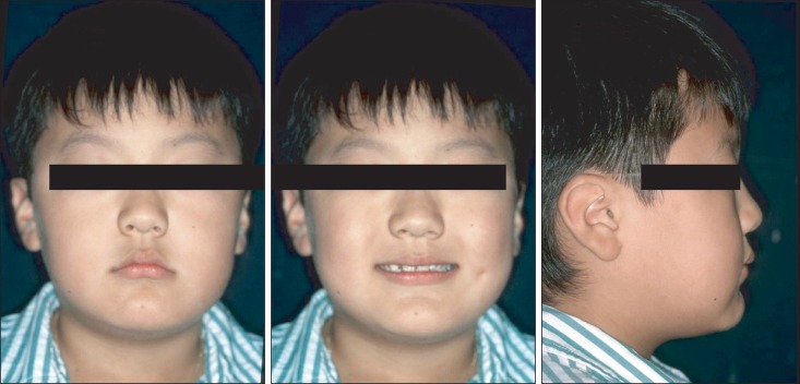
Figure 1 Pretreatment photographs of facial symmetry of the patient at the age of 8 years and 6 months.
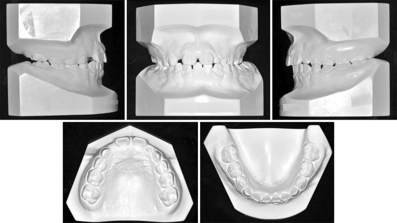
Figure 2 Pretreatment dental casts of the patient at the age of 8 years and 6 months.
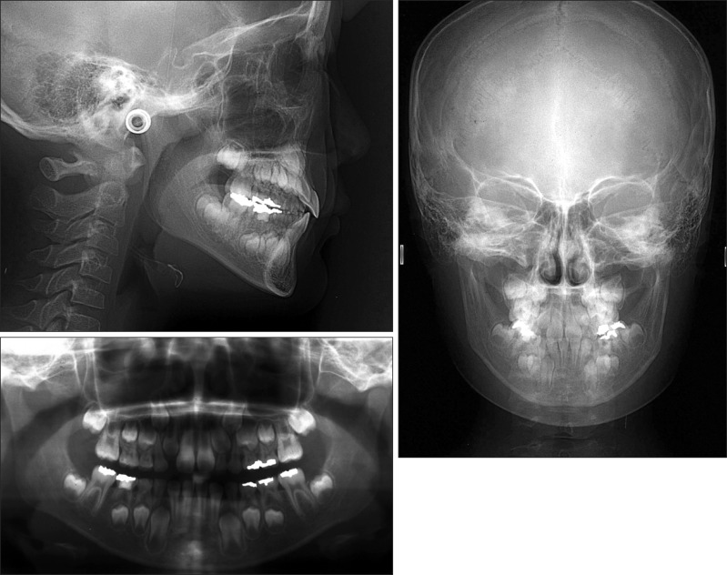
Figure 3 Pretreatment cephalometric and panoramic radiographs obtained at the age of 8 years and 6 months.
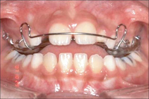
Figure 4 Intraoral photograph of the unilateral posterior crossbite with an expansion plate.
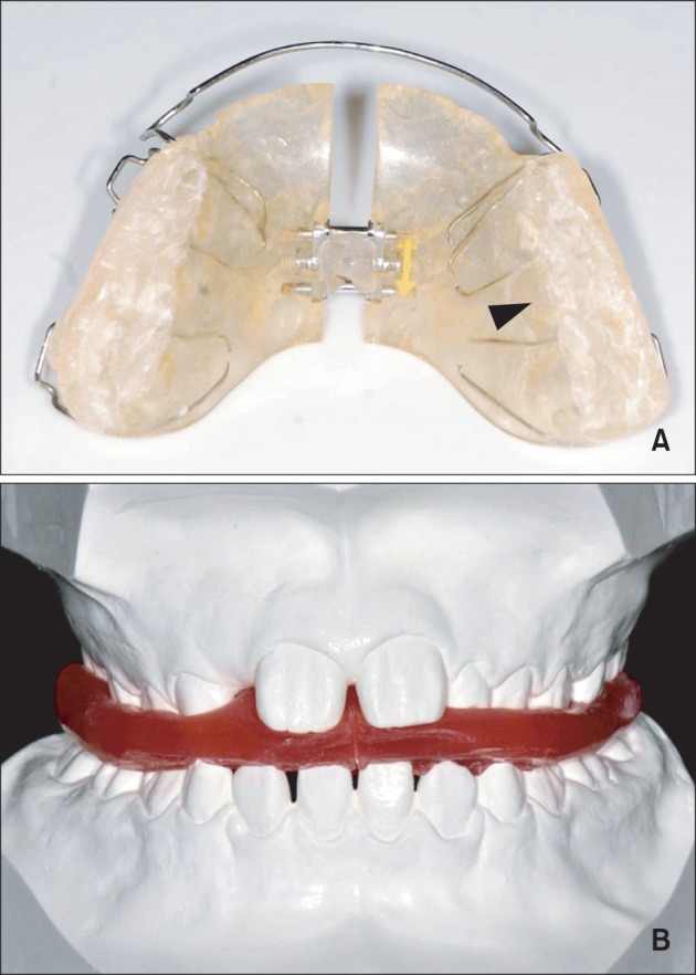
Figure 5 Bite blocks of the expansion plate with occlusal indentations (A, arrow head) were adjusted using the construction bite, which was taken with the incisors in a coincident dental midline (B).
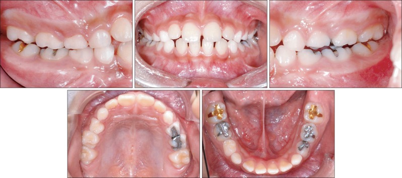
Figure 6 Intraoral photographs obtained 10 months after orthopedic treatment.
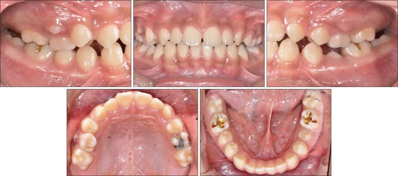
Figure 7 Intraoral photographs obtained after the first phase of treatment (patient age, 10 years and 6 months).
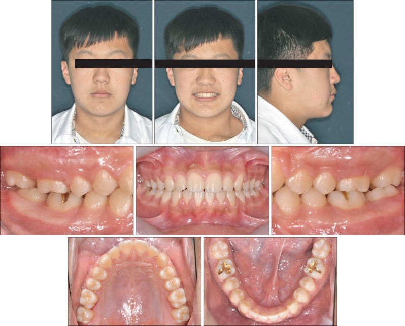
Figure 8 Facial and intraoral photographs obtained after the second phase of treatment (patient age, 13 years and 2 months).

Figure 9 Panoramic and cephalometric radiographs obtained after the second phase of treatment (patient age, 13 years and 2 months).
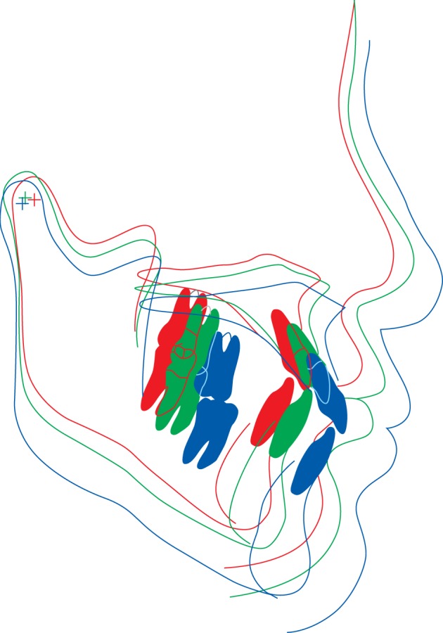
Figure 10 Superimposed cephalometric tracings at pretreatment (red, at 8 years and 6 months of age), after the first phase of treatment (green, at 10 years and 6 months of age), and after the second phase of treatment (blue, at 13 years and 2 months of age).
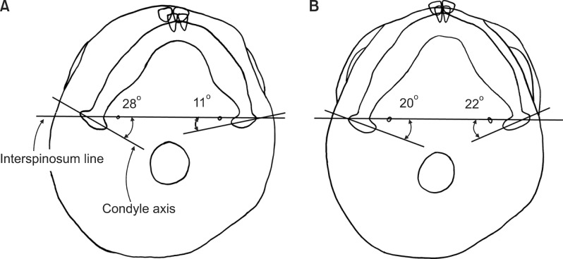
Figure 11 Tracings of submentovertex radiographs. A, Pretreatment (at 8 years and 6 months of age). B, After the second phase of treatment (at 13 years and 2 months of age).
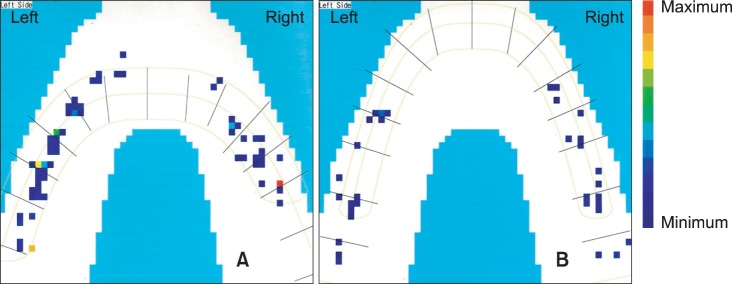
Figure 12 T-scan recordings. A, Pretreatment (at 8 years and 6 months of age). B, After the second phase of treatment (at 13 years and 2 months of age).
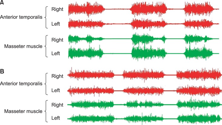
Figure 13 Electromyographic recordings of the anterior temporal muscle and masseter muscle. A, Pretreatment (at 8 years and 6 months of age). B, After the second phase of treatment (at 13 years and 2 months of age).
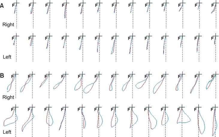
Figure 14 Chewing cycle in the frontal view. The X- and Y-axes represent lateral and vertical movements, respectively. The red line indicates opening and the blue line indicates closing movements. A, Chewing cycles on the right and left side pretreatment. B, Chewing cycles on the right and left side after the second phase of treatment.
Cited by 1 articles
-
Rehabilitation of unstable occlusion caused by inter-dental arch discrepancy
Sun Won, Kiyong An, Chan-Jin Park, Lee-Ra Cho, Yoon-Hyuk Huh
J Korean Acad Prosthodont. 2015;53(4):377-391. doi: 10.4047/jkap.2015.53.4.377.
Reference
-
1. Letzer GM, Kronman JH. A posteroanterior cephalometric evaluation of craniofacial asymmetry. Angle Orthod. 1967; 37:205–211. PMID: 19919216.2. Kutin G, Hawes RR. Posterior cross-bites in the deciduous and mixed dentitions. Am J Orthod. 1969; 56:491–504. PMID: 5261162.
Article3. Egermark-Eriksson I, Carlsson GE, Magnusson T, Thilander B. A longitudinal study on malocclusion in relation to signs and symptoms of cranio-mandibular disorders in children and adolescents. Eur J Orthod. 1990; 12:399–407. PMID: 2086260.
Article4. Schmid W, Mongini F, Felisio A. A computer-based assessment of structural and displacement asymmetries of the mandible. Am J Orthod Dentofacial Orthop. 1991; 100:19–34. PMID: 2069142.
Article5. Andrade AS, Gavião MB, Derossi M, Gameiro GH. Electromyographic activity and thickness of masticatory muscles in children with unilateral posterior crossbite. Clin Anat. 2009; 22:200–206. PMID: 19031391.
Article6. Ingervall B, Thilander B. Activity of temporal and masseter muscles in children with a lateral forced bite. Angle Orthod. 1975; 45:249–258. PMID: 1059337.7. Schröder U, Schröder I. Early treatment of unilateral posterior crossbite in children with bilaterally contracted maxillae. Eur J Orthod. 1984; 6:65–69. PMID: 6583066.8. Hesse KL, Artun J, Joondeph DR, Kennedy DB. Changes in condylar postition and occlusion associated with maxillary expansion for correction of functional unilateral posterior crossbite. Am J Orthod Dentofacial Orthop. 1997; 111:410–418. PMID: 9109586.9. Kecik D, Kocadereli I, Saatci I. Evaluation of the treatment changes of functional posterior crossbite in the mixed dentition. Am J Orthod Dentofacial Orthop. 2007; 131:202–215. PMID: 17276861.
Article10. Cunha RF, Delbem AC, Costa L, de Abreu MG. Treatment of posterior crossbite in mixed dentition with a removable appliance: reports of cases. ASDC J Dent Child. 1999; 66:357–360. PMID: 10631894.11. Garrido García VC, García Cartagena A, González Sequeros O. Evaluation of occlusal contacts in maximum intercuspation using the T-Scan system. J Oral Rehabil. 1997; 24:899–903. PMID: 9467991.
Article12. Melsen B, Stensgaard K, Pedersen J. Sucking habits and their influence on swallowing pattern and prevalence of malocclusion. Eur J Orthod. 1979; 1:271–280. PMID: 298212.
Article13. Harvold EP, Vargervik K, Chierici G. Primate experiments on oral sensation and dental malocclusions. Am J Orthod. 1973; 63:494–508. PMID: 4633053.
Article14. Andrade Ada S, Gameiro GH, Derossi M, Gavião MB. Posterior crossbite and functional changes. A systematic review. Angle Orthod. 2009; 79:380–386. PMID: 19216602.15. Pirttiniemi PM. Associations of mandibular and facial asymmetries--a review. Am J Orthod Dentofacial Orthop. 1994; 106:191–200. PMID: 8059759.
Article16. Sonnesen L, Bakke M. Bite force in children with unilateral crossbite before and after orthodontic treatment. A prospective longitudinal study. Eur J Orthod. 2007; 29:310–313. PMID: 17317867.
Article17. Sultana MH, Yamada K, Hanada K. Changes in occlusal force and occlusal contact area after active orthodontic treatment: a pilot study using pressure-sensitive sheets. J Oral Rehabil. 2002; 29:484–491. PMID: 12028498.
Article18. Ben-Bassat Y, Yaffe A, Brin I, Freeman J, Ehrlich Y. Functional and morphological-occlusal aspects in children treated for unilateral posterior cross-bite. Eur J Orthod. 1993; 15:57–63. PMID: 8436197.
Article19. Brin I, Ben-Bassat Y, Blustein Y, Ehrlich J, Hochman N, Marmary Y, et al. Skeletal and functional effects of treatment for unilateral posterior crossbite. Am J Orthod Dentofacial Orthop. 1996; 109:173–179. PMID: 8638566.
Article
- Full Text Links
-
- Actions
-
Cited
- CITED
-
- Close
- Share
-
- Similar articles
-
- Distraction osteogenesis in facial asymmetry patient
- Level of perception of changed lip protrusion and asymmetry of the lower facial height
- Camouflage treatment of posterior bite collapse in a patient with skeletal asymmetry by using posterior maxillary segmental osteotomy
- Treatment of unilateral buccal crossbite with mandibular symphyseal distraction osteogenesis
- The comparative analysis of the dentition and morphologic malocclusion of the temporomandibular dysfunction patient
