Treatment of unilateral buccal crossbite with mandibular symphyseal distraction osteogenesis
- Affiliations
-
- 1Department of Orthodontics, Faculty of Dentistry, Zonguldak Karaelmas University, Zonguldak, Turkey. dt.nurhat@yahoo.com
- 2Department of Orthodontics, Faculty of Dentistry, Ondokuz Mayis University, Samsun, Turkey.
- 3Department of Oral and Maxillo-facial Surgery, Faculty of Dentistry, Ondokuz Mayis University, Samsun, Turkey.
- KMID: 1762669
- DOI: http://doi.org/10.4041/kjod.2011.41.1.59
Abstract
- The aim of this report is to present the treatment of a 14-year-old boy with scissors-bite. Mandibular symphyseal distraction osteogenesis (MSDO) with tooth-supported distractor was performed to expand the mandible, and intermaxillary cross elastics were used. The mandible was expanded approximately 9 mm. Asymmetric widening was done by using cross elastics and MSDO simultaneously. The buccal crossbite was corrected successfully. After a 2-year observation period, widening of the mandible using this procedure was judged to be stable.
Figure
-
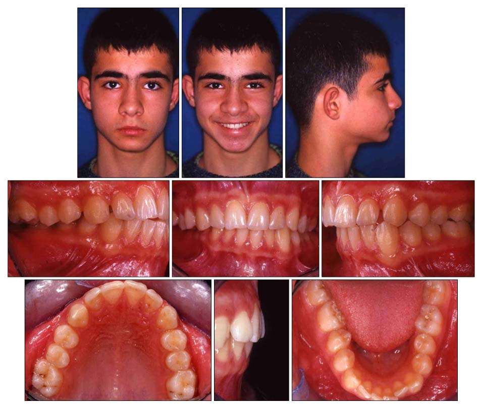
Fig. 1 Pretreatment facial and intraoral photographs.

Fig. 2 Pretreatment panoramic, intraoral periapical (mandibular central incisors), lateral cephalometric, posteroanterior cephalometric, and occlusal mandibular radiographs.
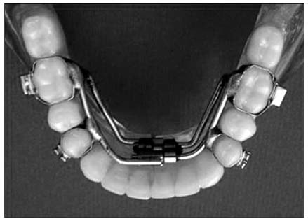
Fig. 3 The tooth-supported distractor. The distractor was fabricated with a hyrax screw, two premolar bands, and two molar bands. The distractor appliance was adjusted to the lower first premolars and the lower first molars.
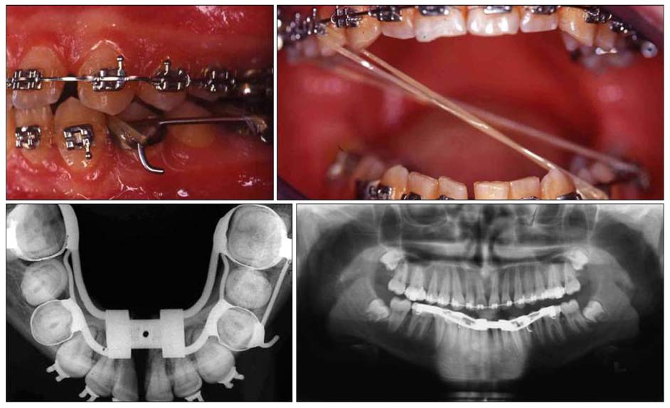
Fig. 4 Occlusal mandibular and panoramic radiographs at the expansion phase of the distraction process and intraoral photographs.
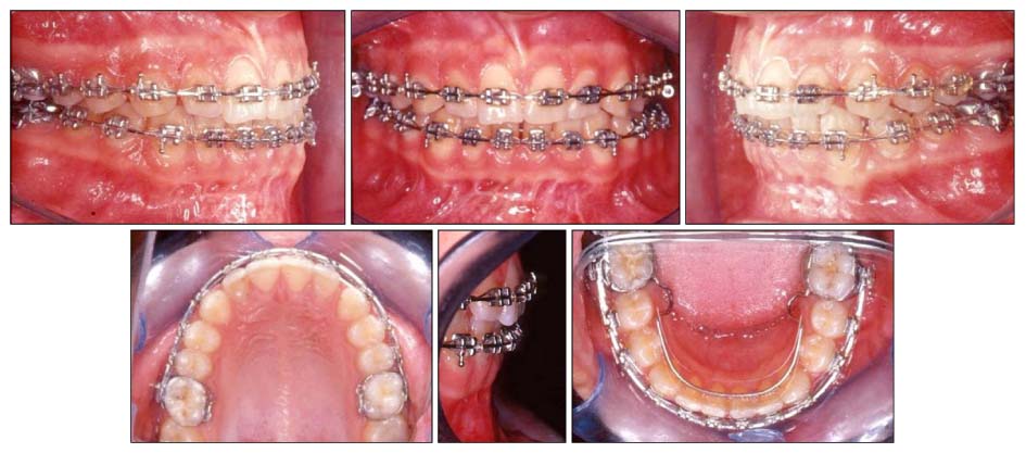
Fig. 5 Postdistraction intraoral photographs. At the end of the 3-month consolidation period, a lingual arch was placed on the lower arch.

Fig. 6 Posttreatment facial and intraoral photographs.

Fig. 7 Posttreatment panoramic, lateral and posteroanterior cephalometric radiographs.
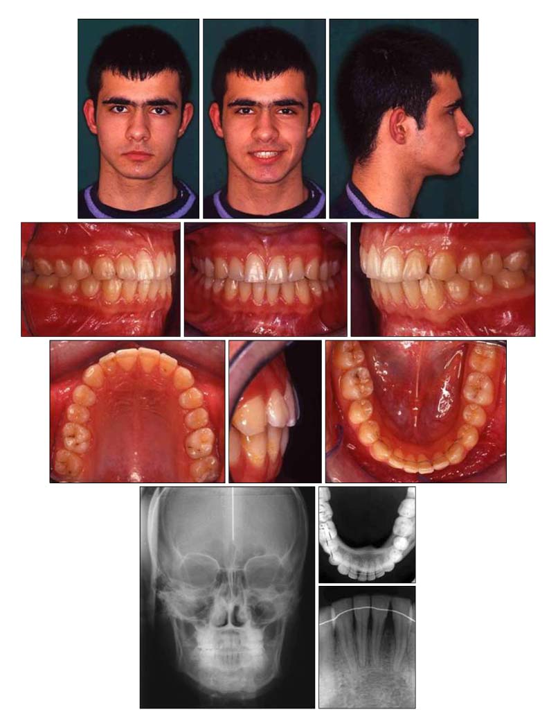
Fig. 8 Postretention facial and intraoral photographs. Posteroanterior cephalometric, occlusal mandibular and intraoral periapical (mandibular central incisors), radiographs.
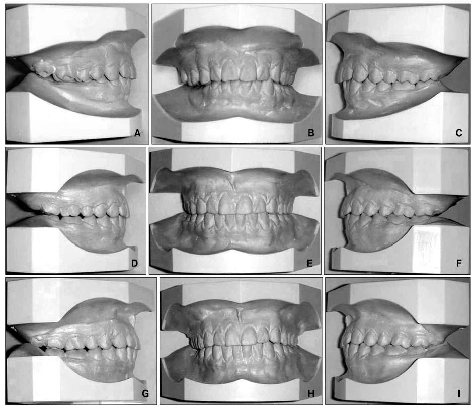
Fig. 9 Photographs of orthodontic models. A-C, Before treatment; D-F, after treatment; G-I, after retention.

Fig. 10 Posteroanterior cephalometric superimposition (― Pretreatment, ― Posttreatment, ― Postretention) and lateral cephalometric superimposition (― Pretreatment, ― Posttreatment, ― Postretention).
Reference
-
1. Tamamura N, Kuroda S, Sugawara Y, Takano-Yamamoto T, Yamashiro T. Use of palatal miniscrew anchorage and lingual multi-bracket appliances to enhance efficiency of molar scissors-bite correction. Angle Orthod. 2009. 79:577–584.
Article2. Bayram M, Ozer M, Alkan A. Mandibular symphyseal distraction osteogenesis using a bone-supported distractor. Angle Orthod. 2007. 77:745–752.
Article3. Yun SW, Lim WH, Chong DR, Chun YS. Scissors-bite correction on second molar with a dragon helix appliance. Am J Orthod Dentofacial Orthop. 2007. 132:842–847.
Article4. Tae KC, Kang KH, Kim SC. Unilateral mandibular widening with distraction osteogenesis. Angle Orthod. 2005. 75:1053–1060.5. Ilizarov GA. The tension-stress effect on the genesis and growth of tissues Part 1. The influence of stability of fixation and soft-tissue preservation. Clin Orthop Relat Res. 1989. (238):249–281.6. Schwarz AM, Gratzinger M. Removable orthodontic appliances. 1966. Philadelphia: WB Saunders;61–83.7. McDougall PD, McNamara JA Jr, Dierkes JM. Arch width development in Class II patients treated with the Fränkel appliance. Am J Orthod. 1982. 82:10–22.
Article8. Herberger RJ. Stability of mandibular intercuspid width after long periods of retention. Angle Orthod. 1981. 51:78–83.9. Blake M, Bibby K. Retention and stability: a review of the literature. Am J Orthod Dentofacial Orthop. 1998. 114:299–306.
Article10. Iseri H, Malkoç S. Long-term skeletal effects of mandibular symphyseal distraction osteogenesis. An implant study. Eur J Orthod. 2005. 27:512–517.
Article11. Little RM, Riedel RA, Stein A. Mandibular arch length increase during the mixed dentition: postretention evaluation of stability and relapse. Am J Orthod Dentofacial Orthop. 1990. 97:393–404.
Article12. Gandini LG Jr, Martins RP, de Oliveira CA, Abdala A, Buschang PH. Midsymphyseal distraction osteogenesis: a new alternative for the treatment of dental crowding. Am J Orthod Dentofacial Orthop. 2009. 135:530–535.
Article13. Conley R, Legan H. Mandibular symphyseal distraction osteogenesis: diagnosis and treatment planning considerations. Angle Orthod. 2003. 73:3–11.14. Samchukov ML, Cope JB, Harper RP, Ross JD. Biomechanical considerations of mandibular lengthening and widening by gradual distraction using a computer model. J Oral Maxillofac Surg. 1998. 56:51–59.
Article15. Cope JB, Harper RP, Samchukov ML. Experimental tooth movement through regenerate alveolar bone: a pilot study. Am J Orthod Dentofacial Orthop. 1999. 116:501–505.
Article16. Mommaerts MY, Polsbroek R, Santler G, Correia PE, Abeloos JV, Ali N. Anterior transmandibular osteodistraction: clinical and model observations. J Craniomaxillofac Surg. 2005. 33:318–325.
Article17. Stelnicki EJ, Stucki-McCormick SU, Rowe N, McCarthy JG. Remodeling of the temporomandibular joint following mandibular distraction osteogenesis in the transverse dimension. Plast Reconstr Surg. 2001. 107:647–658.
Article18. Bell WH, Harper RP, Gonzalez M, Cherkashin AM, Samchukov ML. Distraction osteogenesis to widen the mandible. Br J Oral Maxillofac Surg. 1997. 35:11–19.
Article19. Guerrero CA, Bell WH, Contasti GI, Rodriguez AM. Mandibular widening by intraoral distraction osteogenesis. Br J Oral Maxillofac Surg. 1997. 35:383–392.
Article20. Del Santo M Jr, Guerrero CA, Buschang PH, English JD, Samchukov ML, Bell WH. Long-term skeletal and dental effects of mandibular symphyseal distraction osteogenesis. Am J Orthod Dentofacial Orthop. 2000. 118:485–493.
Article
- Full Text Links
-
- Actions
-
Cited
- CITED
-
- Close
- Share
-
- Similar articles
-
- Clinical study of mandible symphysis widening
- Distraction osteogenesis in collapsed mandible arch patients by accidents
- Distraction osteogenesis on mandible symphysis widening with a bone-borne type distractor
- Reports of mandibular symphysis widening with distraction osteogenesis
- Distraction osteogenesis in patients with hemifacial microsomia
