Korean J Orthod.
2014 May;44(3):128-135. 10.4041/kjod.2014.44.3.128.
Evaluation of the genotoxicity and cytotoxicity in the buccal epithelial cells of patients undergoing orthodontic treatment with three light-cured bonding composites by using micronucleus testing
- Affiliations
-
- 1Department of Orthodontics, Faculty of Dentistry, Inonu University, Malatya, Turkey. ebubekirtoy@gmail.com
- 2Department of Medical Biology and Genetics, Faculty of Medicine, Inonu University, Malatya, Turkey.
- 3Department of Orthodontics, Faculty of Dentistry, Pamukkale University, Denizli, Turkey.
- 4Department of Restorative Dentistry, Faculty of Dentistry, Inonu University, Malatya, Turkey.
- KMID: 1974998
- DOI: http://doi.org/10.4041/kjod.2014.44.3.128
Abstract
OBJECTIVE
This study evaluated the cytotoxicity and genotoxicity of fixed orthodontic treatment with three different light-cured orthodontic bonding composites by analyzing micronucleus (MN) formation in the buccal mucosa during a 6-month period.
METHODS
Thirty healthy volunteers were selected from consecutive patients referred for orthodontic treatment. Equilibrium 2 brackets and molar tubes (Dentaurum) were bonded with three different light-cured orthodontic bonding composites-Transbond XT (3M Unitek), Kurasper F (Kuraray Europe), or GrenGloo (Ormco Corporation)- to all teeth in both arches. Exfoliated buccal epithelial cells were scraped from the middle part of the inner cheeks with sterile cement spatulas before treatment and at 1, 3, and 6 months after treatment. MNs and nuclear alterations, such as karyorrhexis (KR), karyolysis (KL), and binucleated cells (BNs), were scored under a light microscope. Repeated measure ANOVA was used to calculate statistical differences in degenerative nuclear abnormalities.
RESULTS
MN rates did not significantly differ among different time points within the same cell type (p > 0.05). In contrast, the number of BNs in buccal epithelial cells significantly increased in all composite groups (p < 0.01, Transbond XT; p < 0.001, Kurasper F and GrenGloo). KL frequency significantly increased between the beginning and end of the study in the Kurasfer F (0.80 +/- 0.79 to 1.90 +/- 1.10; p < 0.05) and GrenGloo (1.30 +/- 1.06 to 2.40 +/- 1.08; p < 0.05) groups.
CONCLUSIONS
After 6 months of fixed orthodontic treatment with different light-cured composites, morphological signs of cytotoxicity were observed but genotoxic effects were absent.
Keyword
Figure
-
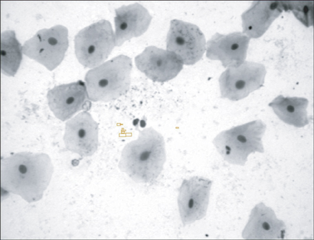
Figure 1 Normal cells stained with acridine orange (×1,000).

Figure 2 Formation of micronucleus stained with acridine orange (×1,000).
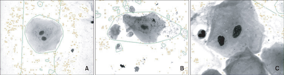
Figure 3 Cytotoxicity evaluation in this study. A, Binucleated cell; B, karyolysis and cell death; C, binucleated cell and karyorrhexis stained with acridine orange (×1,000).
Reference
-
1. Malkoç S, Uysal T, Uşümez S, Işman E, Baysal A. In-vitro assessment of temperature rise in the pulp during orthodontic bonding. Am J Orthod Dentofacial Orthop. 2010; 137:379–383.
Article2. Huang FM, Chou MY, Chang YC. Dentin bonding agents induce c-fos and c-jun protooncogenes expression in human gingival fibroblasts. Biomaterials. 2003; 24:157–163.
Article3. Spahl W, Budzikiewicz H, Geurtsen W. Determination of leachable components from four commercial dental composites by gas and liquid chromatography/mass spectrometry. J Dent. 1998; 26:137–145.
Article4. Söhoel H, Gjerdet NR, Hensten-Pettersen A, Ruyter IE. Allergenic potential of two orthodontic bonding materials. Scand J Dent Res. 1994; 102:126–129.
Article5. Tadin A, Galic N, Mladinic M, Marovic D, Kovacic I, Zeljezic D. Genotoxicity in gingival cells of patients undergoing tooth restoration with two different dental composite materials. Clin Oral Investig. 2014; 18:87–96.
Article6. Koulaouzidou EA, Helvatjoglu-Antoniades M, Palaghias G, Karanika-Kouma A, Antoniades D. Cytotoxicity of dental adhesives in vitro. Eur J Dent. 2009; 3:3–9.
Article7. Ulker HE, Sengun A. Cytotoxicity evaluation of self adhesive composite resin cements by dentin barrier test on 3D pulp cells. Eur J Dent. 2009; 3:120–126.8. Malkoc S, Corekci B, Ulker HE, Yalçin M, Sengün A. Cytotoxic effects of orthodontic composites. Angle Orthod. 2010; 80:571–576.
Article9. Kleinsasser NH, Schmid K, Sassen AW, Harréus UA, Staudenmaier R, Folwaczny M, et al. Cytotoxic and genotoxic effects of resin monomers in human salivary gland tissue and lymphocytes as assessed by the single cell microgel electrophoresis (Comet) assay. Biomaterials. 2006; 27:1762–1770.
Article10. Schweikl H, Schmalz G, Weinmann W. The induction of gene mutations and micronuclei by oxiranes and siloranes in mammalian cells in vitro. J Dent Res. 2004; 83:17–21.
Article11. Öztürk F, Yüksel Ş, Toy E, Kurtoğlu EL, Küçük EB. Genotoxic effects of banding procedure with different orthodontic cements on human oral mucosa cells. Turk J Med Sci. 2012; 42:Suppl 1. 1157–1165.12. Angelieri F, Carlin V, Martins RA, Ribeiro DA. Biomonitoring of mutagenicity and cytotoxicity in patients undergoing fixed orthodontic therapy. Am J Orthod Dentofacial Orthop. 2011; 139:4 Suppl. e399–e404.
Article13. İz SG, Gürhan SİD, Şen BH, Endoğan T, HasircI N. Comparison of in vitro cytotoxicity and genotoxicity of MMA-based polymeric materials and various metallic materials. Turk J Med Sci. 2010; 40:905–916.14. Faccioni F, Franceschetti P, Cerpelloni M, Fracasso ME. In vivo study on metal release from fixed orthodontic appliances and DNA damage in oral mucosa cells. Am J Orthod Dentofacial Orthop. 2003; 124:687–693. discussion 693-4.
Article15. Tolbert PE, Shy CM, Allen JW. Micronuclei and other nuclear anomalies in buccal smears: methods development. Mutat Res. 1992; 271:69–77.
Article16. Gioka C, Bourauel C, Hiskia A, Kletsas D, Eliades T, Eliades G. Light-cured or chemically cured orthodontic adhesive resins? A selection based on the degree of cure, monomer leaching, and cytotoxicity. Am J Orthod Dentofacial Orthop. 2005; 127:413–419. quiz 516.
Article17. Reichl FX, Durner J, Hickel R, Kunzelmann KH, Jewett A, Wang MY, et al. Distribution and excretion of TEGDMA in guinea pigs and mice. J Dent Res. 2001; 80:1412–1415.
Article18. Ağaoğlu G, Arun T, Izgi B, Yarat A. Nickel and chromium levels in the saliva and serum of patients with fixed orthodontic appliances. Angle Orthod. 2001; 71:375–379.19. Kocadereli L, Ataç PA, Kale PS, Ozer D. Salivary nickel and chromium in patients with fixed orthodontic appliances. Angle Orthod. 2000; 70:431–434.20. von der Hude W, Kalweit S, Engelhardt G, McKiernan S, Kasper P, Slacik-Erben R, et al. In vitro micronucleus assay with Chinese hamster V79 cells - results of a collaborative study with in situ exposure to 26 chemical substances. Mutat Res. 2000; 468:137–163.
Article21. Stich HF, Rosin MP. Quantitating the synergistic effect of smoking and alcohol consumption with the micronucleus test on human buccal mucosa cells. Int J Cancer. 1983; 31:305–308.
Article22. Schweikl H, Schmalz G, Spruss T. The induction of micronuclei in vitro by unpolymerized resin monomers. J Dent Res. 2001; 80:1615–1620.
Article23. Santos RL, Pithon MM, Fernandes AB, Cabral MG, Ruellas AC. Biocompatibility of orthodontic adhesives in rat subcutaneous tissue. J Appl Oral Sci. 2010; 18:503–508.
Article24. Bakopoulou A, Mourelatos D, Tsiftsoglou AS, Giassin NP, Mioglou E, Garefis P. Genotoxic and cytotoxic effects of different types of dental cement on normal cultured human lymphocytes. Mutat Res. 2009; 672:103–112.
Article25. Arossi GA, Lehmann M, Dihl RR, Reguly ML, de Andrade HH. Induced DNA damage by dental resin monomers in somatic cells. Basic Clin Pharmacol Toxicol. 2010; 106:124–129.
Article26. Geurtsen W, Lehmann F, Spahl W, Leyhausen G. Cytotoxicity of 35 dental resin composite monomers/additives in permanent 3T3 and three human primary fibroblast cultures. J Biomed Mater Res. 1998; 41:474–480.
Article27. de Soet JJ, Gruythuysen RJ, Bosch JA, van Amerongen WE. The effect of 6-monthly application of 40% chlorhexidine varnish on the microflora and dental caries incidence in a population of children in Surinam. Caries Res. 2002; 36:449–455.
Article28. Baraba A, Zelježić D, Kopjar N, Mladinić M, Anić I, Miletić I. Evaluation of cytotoxic and genotoxic effects of two resin-based root-canal sealers and their components on human leucocytes in vitro. Int Endod J. 2011; 44:652–661.
Article29. Ghose UR, Parida BB. Cytological study of exfoliated buccal mucosa cells of tribes in Orissa State (India) with high risk for oral cancer. Indian J Cancer. 1995; 32:95–99.30. Burgaz S, Işcan A, Büyükbingöl ZK, Bozkurt A, Karakaya AE. Evaluation of micronuclei in exfoliated urothelial cells and urinary thioether excretion of smokers. Mutat Res. 1995; 335:163–169.
Article31. Erdemir EO, Sengün A, Ulker M. Cytotoxicity of mouthrinses on epithelial cells by micronucleus test. Eur J Dent. 2007; 1:80–85.
Article32. Xu GL, Bestor TH, Bourc'his D, Hsieh CL, Tommerup N, Bugge M, et al. Chromosome instability and immunodeficiency syndrome caused by mutations in a DNA methyltransferase gene. Nature. 1999; 402:187–191.
Article33. Cerqueira EM, Meireles JR, Lopes MA, Junqueira VC, Gomes-Filho IS, Trindade S, et al. Genotoxic effects of X-rays on keratinized mucosa cells during panoramic dental radiography. Dentomaxillofac Radiol. 2008; 37:398–403.
Article34. Angelieri F, Carlin V, Saez DM, Pozzi R, Ribeiro DA. Mutagenicity and cytotoxicity assessment in patients undergoing orthodontic radiographs. Dentomaxillofac Radiol. 2010; 39:437–440.
Article35. Moore LE, Warner ML, Smith AH, Kalman D, Smith MT. Use of the fluorescent micronucleus assay to detect the genotoxic effects of radiation and arsenic exposure in exfoliated human epithelial cells. Environ Mol Mutagen. 1996; 27:176–184.
Article
- Full Text Links
-
- Actions
-
Cited
- CITED
-
- Close
- Share
-
- Similar articles
-
- The effects of fluoride releasing orthodontic sealant on the shear bond strength of light-and chemical-cured orthodontic resins
- Shear bond strength of metal brackets bonded with light-cured adhesive: an in vitro comparative study
- In vitro and in vivo evaluation of the genotoxicity of titanium dioxide, GST
- Genotoxicity Assessment of Erythritol by Using Short-term Assay
- Shear bond strength and failure patterns according to the material of resin base in indirect racket bonding
