Comparison of occlusal contact areas of class I and class II molar relationships at finishing using three-dimensional digital models
- Affiliations
-
- 1Department of Dentistry, Graduate School of Medicine, Ewha Womans University, Seoul, Korea. Yschun@ewha.ac.kr
- KMID: 1974802
- DOI: http://doi.org/10.4041/kjod.2015.45.3.113
Abstract
OBJECTIVE
This study compared occlusal contact areas of ideally planned set-up and accomplished final models against the initial in class I and II molar relationships at finishing.
METHODS
Evaluations were performed for 41 post-orthodontic treatment cases, of which 22 were clinically diagnosed as class I and the remainder were diagnosed as full cusp class II. Class I cases had four first premolars extracted, while class II cases had maxillary first premolars extracted. Occlusal contact areas were measured using a three-dimensional scanner and RapidForm 2004. Independent t-tests were used to validate comparison values between class I and II finishings. Repeated measures analysis of variance was used to compare initial, set up, and final models.
RESULTS
Molars from cases in the class I finishing for the set-up model showed significantly greater contact areas than those from class II finishing (p < 0.05). The final model class I finishing showed significantly larger contact areas for the second molars (p < 0.05). The first molars of the class I finishing for the final model showed a tendency to have larger contact areas than those of class II finishing, although the difference was not statistically significant (p = 0.078).
CONCLUSIONS
In set-up models, posterior occlusal contact was better in class I than in class II finishing. In final models, class I finishing tended to have larger occlusal contact areas than class II finishing.
Keyword
MeSH Terms
Figure
-

Figure 1 Classification of class I and II finishings. A, Class I finishing viewed from the buccal aspect. B, Class I finishing viewed from the lingual aspect. C, Class II finishing viewed from the buccal aspect. D, Class II finishing viewed from the lingual aspect.

Figure 2 Calculation of occlusal contact areas of set-up models using the Boolean function (RapidForm 2004) in mm2 areas. A, Occlusal contact area of class I finishing. B, Occlusal contact area of class II finishing.
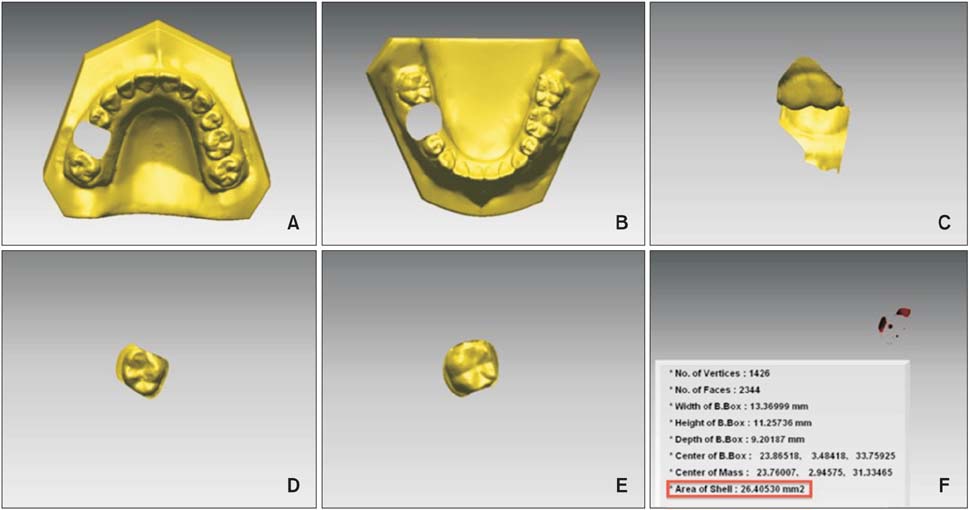
Figure 3 Images of the RapidForm 2004 used to calculate the occlusal contact areas. A, Image of the maxillary model sample with the first molar excised. B, Image of the excised maxillary first molar. C, Buccal aspect of the occluded first molars. D, Image of the excised mandibular first molar. E, Image of the mandibular model sample with the first molar excised. F, Image and measurement of the occlusal contact point (boxed in red).
Cited by 2 articles
-
Comparison of masticatory efficiency according to Angle's classification of malocclusion
Jungin Bae, Woo-Sung Son, Seong-Sik Kim, Soo-Byung Park, Yong-Il Kim
Korean J Orthod. 2017;47(3):151-157. doi: 10.4041/kjod.2017.47.3.151.Posterior dental compensation and occlusal function in adults with different sagittal skeletal malocclusions
Soonshin Hwang, Yoon Jeong Choi, Sooin Jung, Sujin Kim, Chooryung J. Chung, Kyung-Ho Kim
Korean J Orthod. 2020;50(2):98-107. doi: 10.4041/kjod.2020.50.2.98.
Reference
-
1. Liu D, Melsen B. Reappraisal of Class II molar relationships diagnosed from the lingual side. Clin Orthod Res. 2001; 4:97–104.
Article2. Wheeler RC. An atlas of tooth form. Philadelphia: WB Saunders;1969.3. Casko JS, Vaden JL, Kokich VG, Damone J, James RD, Cangialosi TJ, et al. Objective grading system for dental casts and panoramic radiographs. American Board of Orthodontics. Am J Orthod Dentofacial Orthop. 1998; 114:589–599.4. Fotis V, Melsen B, Williams S. Posttreatment changes of skeletal morphology following treatment aimed at restriction of maxillary growth. Am J Orthod. 1985; 88:288–296.
Article5. Jang SY, Kim M, Chun YS. Differences in molar relationships and occlusal contact areas evaluated from the buccal and lingual aspects using 3-dimensional digital models. Korean J Orthod. 2012; 42:182–189.
Article6. Gibbs CH, Mahan PE, Lundeen HC, Brehnan K, Walsh EK, Holbrook WB. Occlusal forces during chewing and swallowing as measured by sound transmission. J Prosthet Dent. 1981; 46:443–449.
Article7. Yoon HR, Choi YJ, Kim KH, Chung C. Comparisons of occlusal force according to occlusal relationship, skeletal pattern, age and gender in Koreans. Korean J Orthod. 2010; 40:304–313.
Article8. Yurkstas A, Manly RS. Measurement of occlusal contact area effective in mastication. Am J Orthod. 1949; 35:185–195.
Article9. Garrido García VC, García Cartagena A, González Sequeros O. Evaluation of occlusal contacts in maximum intercuspation using the T-Scan system. J Oral Rehabil. 1997; 24:899–903.
Article10. Angle EH. Classification of malocclusion. Dental Cosmos. 1899; 41:248–264. 350–357.11. Hellman M. An interpretation of angle's Classification of malocclusion of the teeth supported by the evidence from comparative anatomy and evolution. Dental Cosmos. 1920; 62:476.12. Jang JM, Lee SB. A qualitative and quantitative study on occlusal conditions in health volunteers and athletes with normal occlusions. J Adv Prosthodont. 1998; 36:302–322.13. Ehrlich J, Taicher S. Intercuspal contacts of the natural dentition in centric occlusion. J Prosthet Dent. 1981; 45:419–421.
Article14. Delong R, Ko CC, Anderson GC, Hodges JS, Douglas WH. Comparing maximum intercuspal contacts of virtual dental patients and mounted dental casts. J Prosthet Dent. 2002; 88:622–630.
Article15. Kim JH, Kim KB, Kim WC, Kim JH, Kim HY. Accuracy and precision of polyurethane dental arch models fabricated using a three-dimensional subtractive rapid prototyping method with an intraoral scanning technique. Korean J Orthod. 2014; 44:69–76.
Article
- Full Text Links
-
- Actions
-
Cited
- CITED
-
- Close
- Share
-
- Similar articles
-
- Differences in molar relationships and occlusal contact areas evaluated from the buccal and lingual aspects using 3-dimensional digital models
- A study on the second molar which was malpositioned after orthodontic treatment
- Full mouth rehabilitation of skeletal class II patient with regaining occlusal vertical dimension: a case report
- A study of the etiology of unilateral Class II, division 1 malocclusion
- Posterior dental compensation and occlusal function in adults with different sagittal skeletal malocclusions
