Korean J Orthod.
2020 Mar;50(2):98-107. 10.4041/kjod.2020.50.2.98.
Posterior dental compensation and occlusal function in adults with different sagittal skeletal malocclusions
- Affiliations
-
- 1Department of Orthodontics, Gangnam Severance Dental Hospital, College of Dentistry, Yonsei University, Seoul, Korea. khkim@yuhs.ac
- 2Department of Orthodontics and Institute of Craniofacial Deformity, College of Dentistry, Yonsei University, Seoul, Korea.
- KMID: 2471870
- DOI: http://doi.org/10.4041/kjod.2020.50.2.98
Abstract
OBJECTIVE
The aim of this study was to compare posterior tooth inclinations, occlusal force, and contact area of adults with different sagittal malocclusions.
METHODS
Transverse skeletal parameters and posterior tooth inclinations were evaluated using cone beam computed tomography images, and occlusal force as well as contact area were assessed using pressure-sensitive films in 124 normodivergent adults. A linear mixed model was used to cluster posterior teeth into maxillary premolar, maxillary molar, mandibular premolar, and mandibular molar groups. Differences among Class I, II, and III groups were compared using an analysis of variance test and least significant difference post-hoc test. Correlations of posterior dental inclinations to occlusal function were analyzed using Pearson's correlation analysis.
RESULTS
In male subjects, maxillary premolars and molars had the smallest inclinations in the Class II group while maxillary molars had the greatest inclinations in the Class III group. In female subjects, maxillary molars had the smallest inclinations in the Class II group, while maxillary premolars and molars had the greatest inclinations in the Class III group. Occlusal force and contact area were not significantly different among Class I, II, and III groups.
CONCLUSIONS
Premolar and molar inclinations showed compensatory inclinations to overcome anteroposterior skeletal discrepancy in the Class II and III groups; however, their occlusal force and contact area were similar to those of Class I group. In subjects with normodivergent facial patterns, although posterior tooth inclinations may vary, difference in occlusal function may be clinically insignificant in adults with Class I, II, and III malocclusions.
MeSH Terms
Figure
-
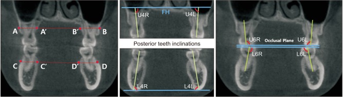
Figure 1 Transverse width and posterior inclination measurements.A–B distance: buccal maxillary width at the midroot level. C–D distance: buccal mandibular width at the midroot level. Maxillary alveolar width: difference between A–B and A′–B′ width divided by 2. Mandibular alveolar width: difference between C–D and C′–D′ width divided by 2. Angle U4R and angle U6R: maxillary right premolar/molar inclination. Angle U4L and angle U6L: maxillary left premolar/molar inclination. Angle L4R and angle L6R: mandibular right premolar/molar inclination. Angle L4L and angle L6L: mandibular left premolar/molar inclination.FH, Frankfort horizontal.
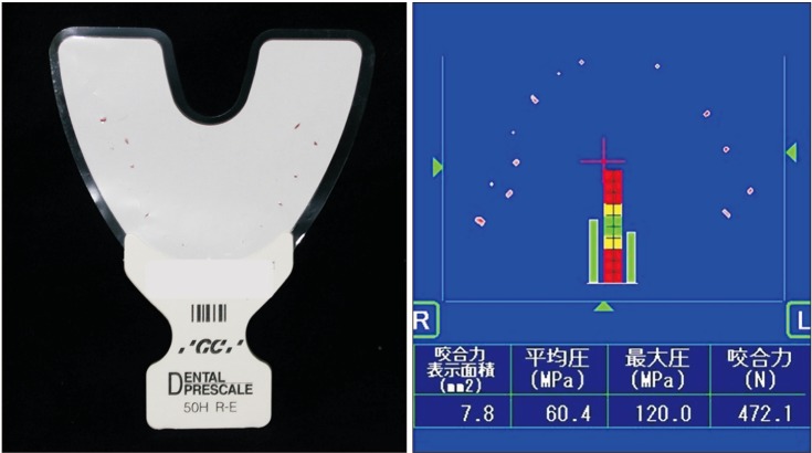
Figure 2 Image of the pressure-sensitive film and CCD camera screen used in the dental prescale system.
Reference
-
1. Angle EH. Treatment of malocclusion of the teeth: angle's system. Philadelphia: White Dental Manufacturing Company;1907.2. Hwang S, Song J, Lee J, Choi YJ, Chung CJ, Kim KH. Three-dimensional evaluation of dentofacial transverse widths in adults with different sagittal facial patterns. Am J Orthod Dentofacial Orthop. 2018; 154:365–374. PMID: 30173839.
Article3. Verma TP, Kumathalli KI, Jain V, Kumar R. Bite force recording devices - a review. J Clin Diagn Res. 2017; 11:ZE01–ZE05.
Article4. Choi YJ, Chung CJ, Kim KH. Changes in occlusal force and occlusal contact area after orthodontic treatment. Korean J Orthod. 2010; 40:176–183.
Article5. Sultana MH, Yamada K, Hanada K. Changes in occlusal force and occlusal contact area after active orthodontic treatment: a pilot study using pressure-sensitive sheets. J Oral Rehabil. 2002; 29:484–491. PMID: 12028498.
Article6. Suzuki T, Kumagai H, Watanabe T, Uchida T, Nagao M. Evaluation of complete denture occlusal contacts using pressure-sensitive sheets. Int J Prosthodont. 1997; 10:386–391. PMID: 9484050.7. Okiyama S, Ikebe K, Nokubi T. Association between masticatory performance and maximal occlusal force in young men. J Oral Rehabil. 2003; 30:278–282. PMID: 12588500.
Article8. Proffit WR, Fields HW, Nixon WL. Occlusal forces in normal- and long-face adults. J Dent Res. 1983; 62:566–570. PMID: 6573373.
Article9. Riedel RA. The relation of maxillary structures to cranium in malocclusion and in normal occlusion. Angle Orthod. 1952; 22:142–145.10. Ahn J, Kim SJ, Lee JY, Chung CJ, Kim KH. Transverse dental compensation in relation to sagittal and transverse skeletal discrepancies in skeletal Class III patients. Am J Orthod Dentofacial Orthop. 2017; 151:148–156. PMID: 28024769.11. Kitafusa Y. Application of “prescale” as an aid to clinical diagnosis in orthodontics. Bull Tokyo Dent Coll. 2004; 45:99–108. PMID: 15536861.
Article12. Ross IF. Occlusal contacts of the natural teeth. J Prosthet Dent. 1974; 32:660–667. PMID: 4530104.
Article13. Braun S, Bantleon HP, Hnat WP, Freudenthaler JW, Marcotte MR, Johnson BE. A study of bite force, part 2: relationship to various cephalometric measurements. Angle Orthod. 1995; 65:373–377. PMID: 8526297.14. Slaj M, Spalj S, Pavlin D, Illes D, Slaj M. Dental archforms in dentoalveolar Class I, II and III. Angle Orthod. 2010; 80:919–924. PMID: 20578864.
Article15. Lux CJ, Conradt C, Burden D, Komposch G. Dental arch widths and mandibular-maxillary base widths in Class II malocclusions between early mixed and permanent dentitions. Angle Orthod. 2003; 73:674–685. PMID: 14719732.16. Baccetti T, Franchi L, McNamara JA Jr, Tollaro I. Early dentofacial features of Class II malocclusion: a longitudinal study from the deciduous through the mixed dentition. Am J Orthod Dentofacial Orthop. 1997; 111:502–509. PMID: 9155809.
Article17. Franchi L, Baccetti T. Transverse maxillary deficiency in Class II and Class III malocclusions: a cephalometric and morphometric study on postero-anterior films. Orthod Craniofac Res. 2005; 8:21–28. PMID: 15667642.
Article18. Wang YL, Cheng J, Chen YM, Yip KH, Smales RJ, Yin XM. Patterns and forces of occlusal contacts during lateral excursions recorded by the T-Scan II system in young Chinese adults with normal occlusions. J Oral Rehabil. 2011; 38:571–578. PMID: 21294761.
Article19. Owens S, Buschang PH, Throckmorton GS, Palmer L, English J. Masticatory performance and areas of occlusal contact and near contact in subjects with normal occlusion and malocclusion. Am J Orthod Dentofacial Orthop. 2002; 121:602–609. PMID: 12080313.
Article20. Lee H, Kim M, Chun YS. Comparison of occlusal contact areas of class I and class II molar relationships at finishing using three-dimensional digital models. Korean J Orthod. 2015; 45:113–120. PMID: 26023539.
Article21. Yoon W, Hwang S, Chung C, Kim KH. Changes in occlusal function after extraction of premolars: 2-year follow-up. Angle Orthod. 2017; 87:703–708. PMID: 28485615.
Article22. Ando K, Fuwa Y, Kurosawa M, Kondo T, Goto S. Bite force measurement system using pressure-sensitive sheet and silicone impression material. Dent Mater J. 2009; 28:212–218. PMID: 19496402.
Article23. Koc D, Dogan A, Bek B. Bite force and influential factors on bite force measurements: a literature review. Eur J Dent. 2010; 4:223–232. PMID: 20396457.
Article24. Maness WL, Podoloff R. Distribution of occlusal contacts in maximum intercuspation. J Prosthet Dent. 1989; 62:238–242. PMID: 2760866.
Article25. Bakke M. Bite force and occlusion. Semin Orthod. 2006; 12:120–126.
Article26. Roldán SI, Restrepo LG, Isaza JF, Vélez LG, Buschang PH. Are maximum bite forces of subjects 7 to 17 years of age related to malocclusion? Angle Orthod. 2016; 86:456–461. PMID: 26347948.
Article27. English JD, Buschang PH, Throckmorton GS. Does malocclusion affect masticatory performance? Angle Orthod. 2002; 72:21–27. PMID: 11843269.28. Toro A, Buschang PH, Throckmorton G, Roldán S. Masticatory performance in children and adolescents with Class I and II malocclusions. Eur J Orthod. 2006; 28:112–119. PMID: 16272209.
Article
- Full Text Links
-
- Actions
-
Cited
- CITED
-
- Close
- Share
-
- Similar articles
-
- The relationship between posterior dental compensation and skeletal discrepancy in class III malocclusion
- Dentoalveolar compensation of hyperdivergent skeletal pattern in children and adults
- Compensatory changes of occlusal plane angles in relation to skeletal factors
- A study on the occlusal plane inclination in lateral cephalograph
- Dentoalveolar Compensation according to Skeletal Patterns of Normal Occlusion
