The effect of radiographic imaging modalities and the observer's experience on postoperative maxillary cyst assessment
- Affiliations
-
- 1Department of Oral and Maxillofacial Radiology and Dental Research Institute, School of Dentistry, Seoul National University, Seoul, Korea. raychoi@snu.ac.kr
- KMID: 1974499
- DOI: http://doi.org/10.5624/isd.2014.44.4.301
Abstract
- PURPOSE
The purpose of this study was to compare the accuracy of postoperative maxillary cyst (POMC) diagnosis by panoramic radiographs versus computed tomography (CT) and by oral and maxillofacial radiologists versus non-specialists.
MATERIALS AND METHODS
Sixty-five maxillary sinuses with POMCs and 63 without any lesion were assessed using panoramic radiographs and CT images by five oral and maxillofacial radiologists and five non-specialists on a five-point scale. The areas under receiver operating characteristic (ROC) curves were analyzed to determine the differences in diagnostic accuracy between the two imaging modalities and between the two groups of observers. The intra-observer agreement was determined, too.
RESULTS
The diagnostic accuracy of CT images was higher than that of panoramic radiographs in both groups of observers (p<0.05). The diagnostic accuracy of oral and maxillofacial radiologists for each method was higher than that of non-specialists (p<0.05).
CONCLUSION
The use of CT improves the diagnosis of POMC, and radiological training and experience leads to more accurate evaluation.
Keyword
MeSH Terms
Figure
-
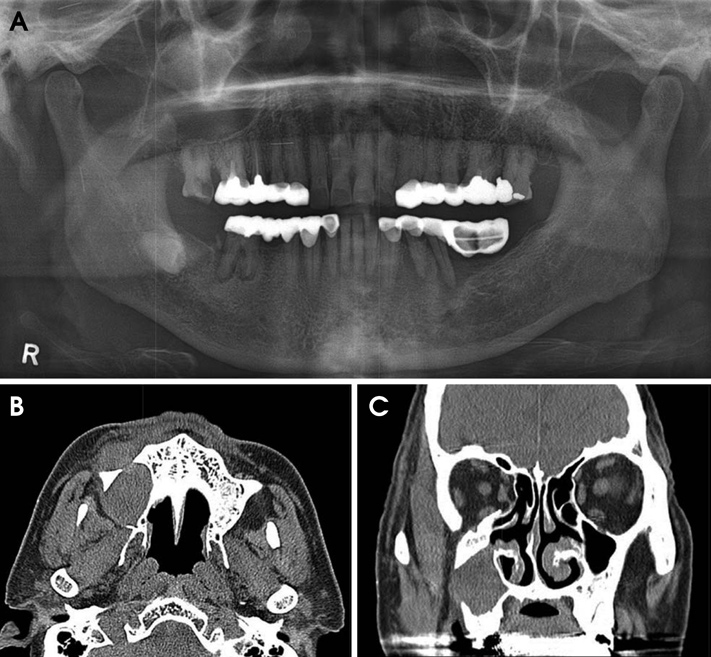
Fig. 1 A. Panoramic radiograph shows a postoperative maxillary cyst in the right maxillary sinus involving the inferior and posterolateral walls. B. An axial computed tomography (CT) image shows an expansile lesion with the anterior and posterolateral wall perforation of the maxillary sinus. C. A coronal CT image shows the lateral wall perforation of the right maxillary sinus.
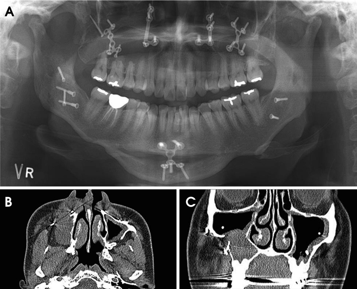
Fig. 2 A. Panoramic radiograph shows a postoperative maxillary cyst in the right maxillary sinus due to maxillary orthognathic surgery. B. An axial CT image shows an expansile lesion with the anterior and posterolateral wall perforation in the maxillary sinus. C. A coronal CT image shows the lateral wall perforation and surgical screw with the right maxillary sinus.
Cited by 1 articles
-
Dental students' ability to detect maxillary sinus abnormalities: A comparison between panoramic radiography and cone-beam computed tomography
Lucas de Paula Lopes Rosado, Izabele Sales Barbosa, Sibele Nascimento de Aquino, Rafael Binato Junqueira, Francielle Silvestre Verner
Imaging Sci Dent. 2019;49(3):191-199. doi: 10.5624/isd.2019.49.3.191.
Reference
-
1. Yamamoto H, Takagi M. Clinicopathologic study of the postoperative maxillary cyst. Oral Surg Oral Med Oral Pathol. 1986; 62:544–548.
Article2. Kaneshiro S, Nakajima T, Yoshikawa Y, Iwasaki H, Tokiwa N. The postoperative maxillary cyst: report of 71 cases. J Oral Surg. 1981; 39:191–198.3. Lee GI, Park TW. Clinical and radiological study of the postoperative maxillary cyst. J Korean Acad Oral Maxillofac Radiol. 1994; 24:47–55.4. Lyon HE. Reliability of panoramic radiography in the diagnosis of maxillary sinus pathosis. Oral Surg Oral Med Oral Pathol. 1973; 35:124–128.
Article5. Moilanen A. Panoramic zonography in the diagnosis of the maxillary sinus disease. Int J Oral Surg. 1984; 13:432–436.
Article6. Pe MB, Sano K, Kitamura A, Inokuchi T. Computed tomography in the evaluation of postoperative maxillary cysts. J Oral Maxillofac Surg. 1990; 48:679–684.
Article7. Isoda H, Takehara Y, Masui T, Seki A, Takahashi M, Kaneko M. MRI of postoperative maxillary cysts. J Comput Assist Tomogr. 1993; 17:572–575.
Article8. Chindasombatjaroen J, Uchiyama Y, Kakimoto N, Murakami S, Furukawa S, Kishino M. Postoperative maxillary cysts: magnetic resonance imaging compared with computerized tomography. Oral Surg Oral Med Oral Pathol Oral Radiol Endod. 2009; 107:e38–e44.
Article9. Park SH, Goo JM, Jo CH. Receiver operating characteristic (ROC) curve: practical review for radiologists. Korean J Radiol. 2004; 5:11–18.
Article10. Basu MK, Rout PG, Rippin JW, Smith AJ. The post-operative maxillary cyst. Experience with 23 cases. Int J Oral Maxillofac Surg. 1988; 17:282–284.11. Sugar AW, Walker DM, Bounds GA. Surgical ciliated (postoperative maxillary) cysts following mid-face osteotomies. Br J Oral Maxillofac Surg. 1990; 28:264–267.
Article12. Hayhurst DL, Moenning JE, Summerlin DJ, Bussard DA. Surgical ciliated cyst: a delayed complication in a case of maxillary orthognathic surgery. J Oral Maxillofac Surg. 1993; 51:705–709.
Article13. Amin M, Witherow H, Lee R, Blenkinsopp P. Surgical ciliated cyst after maxillary orthognathic surgery: report of a case. J Oral Maxillofac Surg. 2003; 61:138–141.
Article14. Kim JJ, Freire M, Yoon JH, Kim HK. Postoperative maxillary cyst after maxillary sinus augmentation. J Craniofac Surg. 2013; 24:e521–e523.
Article15. Ohba T, Katayama H. Panoramic roentgen anatomy of the maxillary sinus. Oral Surg Oral Med Oral Pathol. 1975; 39:658–664.
Article16. Ohba T, Yang R, Chen C, Ogawa Y, Katayama H. Experimental explanation of maxillary sinus radiopacity as seen by Waters' and panoramic projections. Dentomaxillofac Radiol. 1985; 14:133–136.
Article17. Ohba T, Ogawa Y, Hiromatsu T, Shinohara Y. Experimental comparison of radiographic techniques in the detection of maxillary sinus disease. Dentomaxillofac Radiol. 1990; 19:13–17.
Article18. Heo MS, Song MY, Lee SS, Choi SC, Park TW. A comparative study of the radiological diagnosis of postoperative maxillary cyst. Dentomaxillofac Radiol. 2000; 29:347–351.
Article19. Cable HR, Jeans WD, Cullen RJ, Bull PD, Maw AR. Computerized tomography of the Caldwell-Luc cavity. J Laryngol Otol. 1981; 95:775–783.
Article20. Yoshimura Y, Matsuura R, Santo E. Various appearances of postoperative maxillary cysts with computerized tomography. Dentomaxillofac Radiol. 1985; 14:37–40.
Article
- Full Text Links
-
- Actions
-
Cited
- CITED
-
- Close
- Share
-
- Similar articles
-
- A Postoperative Cheek Cyst Originating from the Maxillary Sinus with Orbital Involvement
- Oroantral communication, its causes, complications, treatments and radiographic features: A pictorial review
- Effect of LCD monitor type and observer experience on diagnostic performance in soft-copy interpretations of the maxillary sinus on panoramic radiographs
- A Case of Secondary Trigeminal Neuralgia Caused by Infected Postoperative Maxillary Cyst
- Clinicopathologic study on cysts and postoperative cyst in maxillary sinus
