Imaging Sci Dent.
2014 Sep;44(3):213-220. 10.5624/isd.2014.44.3.213.
Preoperative implant planning considering alveolar bone grafting needs and complication prediction using panoramic versus CBCT images
- Affiliations
-
- 1OIC, OMFS IMPATH Research Group, Department of Imaging and Pathology, Faculty of Medicine, University of Leuven, Leuven, Belgium. reinhilde.jacobs@uzleuven.be
- 2Master of Periodontology, Universidad San Martin de Porres, Lima, Peru.
- 3Department of Oral and Maxillofacial Surgery, University Hospitals, Leuven, Belgium.
- KMID: 1974485
- DOI: http://doi.org/10.5624/isd.2014.44.3.213
Abstract
- PURPOSE
This study was performed to determine the efficacy of observers' prediction for the need of bone grafting and presence of perioperative complications on the basis of cone-beam computed tomography (CBCT) and panoramic radiographic (PAN) planning as compared to the surgical outcome.
MATERIALS AND METHODS
One hundred and eight partially edentulous patients with a need for implant rehabilitation were referred for preoperative imaging. Imaging consisted of PAN and CBCT images. Four observers carried out implant planning using PAN image datasets, and at least one month later, using CBCT image datasets. Based on their own planning, the observers assessed the need for bone graft augmentation as well as complication prediction. The implant length and diameter, the need for bone graft augmentation, and the occurrence of anatomical complications during planning and implant placement were statistically compared.
RESULTS
In the 108 patients, 365 implants were installed. Receiver operating characteristic analyses of both PAN and CBCT preoperative planning showed that CBCT performed better than PAN-based planning with respect to the need for bone graft augmentation and perioperative complications. The sensitivity and the specificity of CBCT for implant complications were 96.5% and 90.5%, respectively, and for bone graft augmentation, they were 95.2% and 96.3%, respectively. Significant differences were found between PAN-based planning and the surgery of posterior implant lengths.
CONCLUSION
Our findings indicated that CBCT-based preoperative implant planning enabled treatment planning with a higher degree of prediction and agreement as compared to the surgical standard. In PAN-based surgery, the prediction of implant length was poor.
Keyword
MeSH Terms
Figure
-

Fig. 1 Receiver operating characteristic (ROC) analysis reveals an excellent diagnostic accuracy of CBCT for the planning of complications and the diagnosis of bone graft augmentation with an area under the curve (AUC) of 0.94.
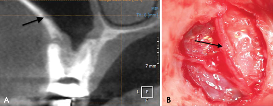
Fig. 2 A. CBCT shows the posterior superior alveolar artery before creating a lateral window into the maxillary sinus. B. The artery is seen during the sinus lift procedure located at 15 mm from the alveolar crest as diagnosed with CBCT.
Cited by 1 articles
-
Analysis of the root position of the maxillary incisors in the alveolar bone using cone-beam computed tomography
Yun-Hoa Jung, Bong-Hae Cho, Jae Joon Hwang
Imaging Sci Dent. 2017;47(3):181-187. doi: 10.5624/isd.2017.47.3.181.
Reference
-
1. Del Fabbro M, Rosano G, Taschieri S. Implant survival rates after maxillary sinus augmentation. Eur J Oral Sci. 2008; 116:497–506.
Article2. Emmerich D, Att W, Stappert C. Sinus floor elevation using osteotomes: a systematic review and meta-analysis. J Periodontol. 2005; 76:1237–1251.
Article3. Pjetursson BE, Tan WC, Zwahlen M, Lang NP. A systematic review of the success of sinus floor elevation and survival of implants inserted in combination with sinus floor elevation. J Clin Periodontol. 2008; 35:216–240.
Article4. Harris D, Horner K, Gröndahl K, Jacobs R, Helmrot E, Benic GI, et al. E.A.O. guidelines for the use of diagnostic imaging in implant dentistry 2011. A consensus workshop organized by the European Association for Osseointegration at the Medical University of Warsaw. Clin Oral Implants Res. 2012; 23:1243–1253.
Article5. Ella B, Sédarat C, Noble Rda C, Normand E, Lauverjat Y, Siberchicot F, et al. Vascular connections of the lateral wall of the sinus: surgical effect in sinus augmentation. Int J Oral Maxillofac Implants. 2008; 23:1047–1052.6. Flanagan D. Arterial supply of maxillary sinus and potential for bleeding complication during lateral approach sinus elevation. Implant Dent. 2005; 14:336–338.
Article7. Kalpidis CD, Setayesh RM. Hemorrhaging associated with endosseous implant placement in the anterior mandible: a review of the literature. J Periodontol. 2004; 75:631–645.
Article8. Greenstein G, Cavallaro J, Romanos G, Tarnow D. Clinical recommendations for avoiding and managing surgical complications associated with implant dentistry: a review. J Periodontol. 2008; 79:1317–1329.
Article9. Misch K, Wang HL. Implant surgery complications: etiology and treatment. Implant Dent. 2008; 17:159–168.
Article10. Jacobs R, Quirynen M, Bornstein M. Neurovascular disturbances after implant surgery. Periodontol 2000. 2014; 66:188–202.
Article11. Guerrero ME, Botetano R, Beltran J, Horner K, Jacobs R. Can preoperative imaging help to predict postoperative outcome after wisdom tooth removal? A randomized controlled trial using panoramic radiography versus cone-beam CT. Clin Oral Investig. 2014; 18:335–342.
Article12. Fleiss JL. Statistical methods for rates and proportions. 2nd ed. New York: Wiley-Interscience;1981. p. 229–232.13. Egger M, Smith GD, Altman DG. Systematic reviews in health care: meta-analysis in context. 2nd ed. London: BMJ;2001. p. 248–282.14. Timmenga NM, Raghoebar GM, Liem RS, van Weissenbruch R, Manson WL, Vissink A. Effects of maxillary sinus floor elevation surgery on maxillary sinus physiology. Eur J Oral Sci. 2003; 111:189–197.
Article15. van den Bergh JP, ten Bruggenkate CM, Disch FJ, Tuinzing DB. Anatomical aspects of sinus floor elevations. Clin Oral Implants Res. 2000; 11:256–265.
Article16. Apostolakis D, Brown JE. The anterior loop of the inferior alveolar nerve: prevalence, measurement of its length and a recommendation for interforaminal implant installation based on cone beam CT imaging. Clin Oral Implants Res. 2012; 23:1022–1030.
Article17. Schropp L, Stavropoulos A, Gotfredsen E, Wenzel A. Comparison of panoramic and conventional cross-sectional tomography for preoperative selection of implant size. Clin Oral Implants Res. 2011; 22:424–429.
Article18. Guerrero ME, Noriega J, Castro C, Jacobs R. Does cone-beam CT alter treatment plans? Comparison of preoperative implant planning using panoramic versus cone-beam CT images. Imaging Sci Dent. 2014; 44:121–128.
Article19. Renton T, Dawood A, Shah A, Searson L, Yilmaz Z. Post-implant neuropathy of the trigeminal nerve. A case series. Br Dent J. 2012; 212:E17.
Article20. Temmerman A, Hertelé S, Teughels W, Dekeyser C, Jacobs R, Quirynen M. Are panoramic images reliable in planning sinus augmentation procedures? Clin Oral Implants Res. 2011; 22:189–194.
Article21. Marx RE, Garg AK. A novel aid to elevation of the sinus membrane for the sinus lift procedure. Implant Dent. 2002; 11:268–271.
Article22. Testori T, Wallace SS, Del Fabbro M, Taschieri S, Trisi P, Capelli M, et al. Repair of large sinus membrane perforations using stabilized collagen barrier membranes: surgical techniques with histologic and radiographic evidence of success. Int J Periodontics Restorative Dent. 2008; 28:9–17.23. Cho SC, Wallace SS, Froum SJ, Tarnow DP. Influence of anatomy on Schneiderian membrane perforations during sinus elevation surgery: three-dimensional analysis. Pract Proced Aesthet Dent. 2001; 13:160–163.24. Braut V, Bornstein MM, Belser U, Buser D. Thickness of the anterior maxillary facial bone wall - a retrospective radiographic study using cone beam computed tomography. Int J Periodontics Restorative Dent. 2011; 31:125–131.25. Chan HL, Garaicoa-Pazmino C, Suarez F, Monje A, Benavides E, Oh TJ, et al. Incidence of implant buccal plate fenestration in the esthetic zone: a cone beam computed tomography study. Int J Oral Maxillofac Implants. 2014; 29:171–177.
Article
- Full Text Links
-
- Actions
-
Cited
- CITED
-
- Close
- Share
-
- Similar articles
-
- Comparing the precision of panoramic radiography and cone-beam computed tomography in avoiding anatomical structures critical to dental implant surgery: A retrospective study
- Does cone-beam CT alter treatment plans? Comparison of preoperative implant planning using panoramic versus cone-beam CT images
- CBCT analysis of three implant cases for treatment planning
- Evaluation of peri-implant bone defects on cone-beam computed tomography and the diagnostic accuracy of detecting these defects on panoramic images
- Comparison of cone-beam computed tomography and digital panoramic radiography for detecting peri-implant alveolar bone changes using trabecular micro-structure analysis
