Comparing the precision of panoramic radiography and cone-beam computed tomography in avoiding anatomical structures critical to dental implant surgery: A retrospective study
- Affiliations
-
- 1Department of Oral and Maxillofacial Surgery, Faculty of Dentistry, Akdeniz University, Antalya, Turkey. oznur_ozalp_@hotmail.com
- 2Department of Prosthodontics, Faculty of Dentistry, Akdeniz University, Antalya, Turkey.
- KMID: 2450167
- DOI: http://doi.org/10.5624/isd.2018.48.4.269
Abstract
- PURPOSE
The aim of this study was to evaluate the correlations between measurements made using panoramic radiography and cone-beam computed tomography (CBCT) based on certain anatomical landmarks of the jaws, with the goal of preventing complications due to inaccurate measurements in the pre-surgical planning phase of dental implant placement.
MATERIALS AND METHODS
A total of 56 individuals who underwent panoramic radiography and a CBCT evaluation before dental implant surgery were enrolled in the study. Measurements were performed to identify the shortest vertical distance between the alveolar crest and neighboring anatomical structures, including the maxillary sinus, nasal floor, mandibular canal, and foramen mentale. The differences between the measurements on panoramic radiography and CBCT images were statistically analyzed.
RESULTS
Statistically significant differences were observed between the measurements on panoramic radiography and CBCT for all anatomical structures (P < .05). The correlation coefficients (r) between the paired samples obtained from panoramic radiography and CBCT were closely correlated (P < .05), with r values varying from 0.921 and 0.979 for different anatomical regions.
CONCLUSION
The results of this study support the idea that panoramic radiography might provide sufficient information on bone height for preoperative implant planning in routine cases or when CBCT is unavailable. However, an additional CBCT evaluation might be helpful in cases where a safety margin cannot be respected due to insufficient bone height.
MeSH Terms
Figure
-
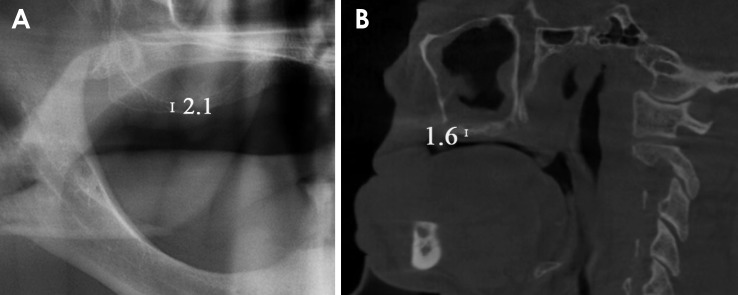
Fig. 1 Measurement of the shortest vertical distance between the alveolar crest and the bottom of the maxillary sinus on a panoramic image (A) and a cone-beam computed tomography image (B).
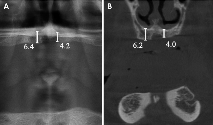
Fig. 2 Measurement of the shortest vertical distance between the alveolar crest and the bottom of the nasal floor on a panoramic image (A) and a cone-beam computed tomography image (B).
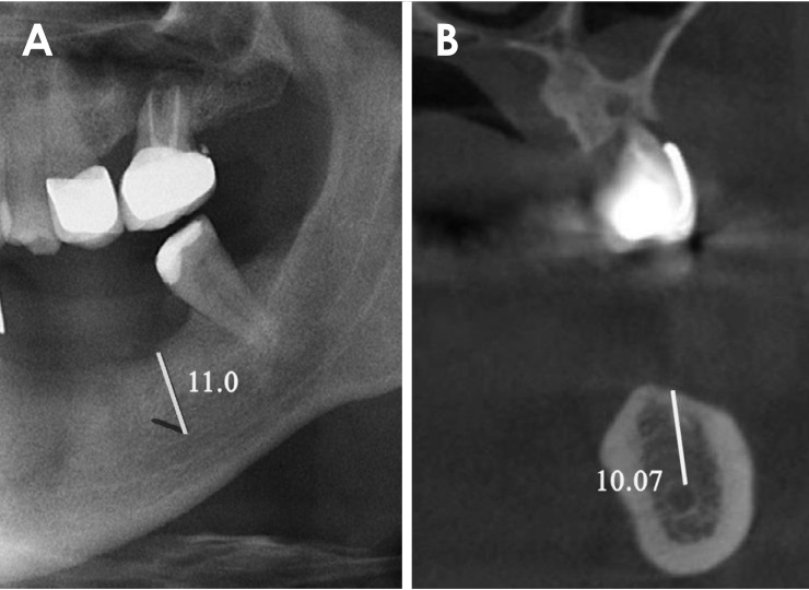
Fig. 3 Measurement of the shortest vertical distance between the alveolar crest and the top of the mandibular canal on a panoramic image (A) and a cone-beam computed tomography image (B).
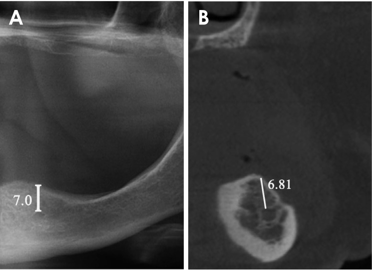
Fig. 4 Measurement of the shortest vertical distance between the alveolar crest and the top of the foramen mentale on a panoramic image (A) and a cone-beam computed tomography image (B).
Cited by 1 articles
-
Cone-beam computed tomography-guided three-dimensional evaluation of treatment effectiveness of the Frog appliance
Mujia Li, Xiaoxia Su, Yang Li, Xianglin Li, Xinqin Si
Korean J Orthod. 2019;49(3):161-169. doi: 10.4041/kjod.2019.49.3.161.
Reference
-
1. Ali SA, Karthigeyan S, Deivanai M, Kumar A. Implant rehabilitation for atrophic maxilla: a review. J Indian Prosthodont Soc. 2014; 14:196–207.
Article2. Misch K, Wang HL. Implant surgery complications: etiology and treatment. Implant Dent. 2008; 17:159–168. PMID: 18545047.
Article3. Jaju PP, Jaju SP. Clinical utility of dental cone-beam computed tomography: current perspectives. Clin Cosmet Investig Dent. 2014; 6:29–43.
Article4. Ahlqwist M, Halling A, Hollender L. Rotational panoramic radiography in epidemiological studies of dental health. Comparison between panoramic radiographs and intraoral full mouth surveys. Swed Dent J. 1986; 10:73–84. PMID: 3518113.5. Lecomber AR, Yoneyama Y, Lovelock DJ, Hosoi T, Adams AM. Comparison of patient dose from imaging protocols for dental implant planning using conventional radiography and computed tomography. Dentomaxillofac Radiol. 2001; 30:255–259. PMID: 11571544.
Article6. Monsour PA, Dudhia R. Implant radiography and radiology. Aust Dent J. 2008; 53(Suppl 1):S11–S25. PMID: 18498579.
Article7. Jacobs R, Quirynen M. Dental cone beam computed tomography: justification for use in planning oral implant placement. Periodontol 2000. 2014; 66:203–213. PMID: 25123769.
Article8. Verstreken K, Van Cleynenbreugel J, Marchal G, Naert I, Suetens P, van Steenberghe D. Computer-assisted planning of oral implant surgery: a three-dimensional approach. Int J Oral Maxillofac Implants. 1996; 11:806–810. PMID: 8990645.9. Amarnath GS, Kumar U, Hilal M, Muddugangadhar BC, Anshuraj K, Shruthi CS. Comparison of cone beam computed tomography, orthopantomography with direct ridge mapping for pre-surgical planning to place implants in cadaveric mandibles: an ex-vivo study. J Int Oral Health. 2015; 7(Suppl 1):38–42.10. Klatt JC, Heiland M, Marx S, Hanken H, Schmelzle R, Pohlenz P. Clinical indication for intraoperative 3D imaging during open reduction of fractures of the mandibular angle. J Craniomaxillofac Surg. 2013; 41:e87–e90. PMID: 23333491.
Article11. Hu KS, Choi DY, Lee WJ, Kim HJ, Jung UW, Kim S. Reliability of two different presurgical preparation methods for implant dentistry based on panoramic radiography and cone-beam computed tomography in cadavers. J Periodontal Implant Sci. 2012; 42:39–44. PMID: 22586521.
Article12. Pertl L, Gashi-Cenkoglu B, Reichmann J, Jakse N, Pertl C. Preoperative assessment of the mandibular canal in implant surgery: comparison of rotational panoramic radiography (OPG), computed tomography (CT) and cone beam computed tomography (CBCT) for preoperative assessment in implant surgery. Eur J Oral Implantol. 2013; 6:73–80. PMID: 23513204.13. Correa LR, Spin-Neto R, Stavropoulos A, Schropp L, da Silveira HE, Wenzel A. Planning of dental implant size with digital panoramic radiographs, CBCT-generated panoramic images, and CBCT cross-sectional images. Clin Oral Implants Res. 2014; 25:690–695. PMID: 23442085.
Article14. Koo TK, Li MY. A guideline of selecting and reporting intraclass correlation coefficients for reliability research. J Chiropr Med. 2016; 15:155–163. PMID: 27330520.
Article15. Corpas Ldos S, Jacobs R, Quirynen M, Huang Y, Naert I, Duyck J. Peri-implant bone tissue assessment by comparing the outcome of intra-oral radiograph and cone beam computed tomography analyses to the histological standard. Clin Oral Implants Res. 2011; 22:492–499. PMID: 21143531.16. Isidor F. Clinical probing and radiographic assessment in relation to the histologic bone level at oral implants in monkeys. Clin Oral Implants Res. 1997; 8:255–264. PMID: 9586471.17. Greenstein G, Cavallaro J, Romanos G, Tarnow D. Clinical recommendations for avoiding and managing surgical complications associated with implant dentistry: a review. J Periodontol. 2008; 79:1317–1329. PMID: 18672980.
Article18. Charyeva O, Altynbekov K, Zhartybaev R, Sabdanaliev A. Long-term dental implant success and survival--a clinical study after an observation period up to 6 years. Swed Dent J. 2012; 36:1–6. PMID: 22611899.19. Ritter L, Elger M, Rothamel D, Fienitz T, Zinser M, Schwarz F, et al. Accuracy of peri-implant bone evaluation using cone beam CT, digital intra-oral radiographs and histology. Dentomaxillofac Radiol. 2014; 43:20130088. PMID: 24786136.
Article20. Suomalainen A, Pakbaznejad Esmaeili E, Robinson S. Dentomaxillofacial imaging with panoramic views and cone beam CT. Insights Imaging. 2015; 6:1–16.
Article21. Tang Z, Liu X, Chen K. Comparison of digital panoramic radiography versus cone beam computerized tomography for measuring alveolar bone. Head Face Med. 2017; 13:2. PMID: 28228135.
Article22. Laster WS, Ludlow JB, Bailey LJ, Hershey HG. Accuracy of measurements of mandibular anatomy and prediction of asymmetry in panoramic radiographic images. Dentomaxillofac Radiol. 2005; 34:343–349. PMID: 16227476.
Article23. Choi JW. Assessment of panoramic radiography as a national oral examination tool: review of the literature. Imaging Sci Dent. 2011; 41:1–6. PMID: 21977466.
Article24. Hassan B, Jacobs R. Cone beam computed tomography - 3D imaging in oral and maxillofacial surgery. Eur Med Imaging Rev. 2008; 1:38–40.25. Dreiseidler T, Mischkowski RA, Neugebauer J, Ritter L, Zöller JE. Comparison of cone-beam imaging with orthopantomography and computerized tomography for assessment in presurgical implant dentistry. Int J Oral Maxillofac Implants. 2009; 24:216–225. PMID: 19492636.26. Sheikhi M, Dakhil-Alian M, Bahreinian Z. Accuracy and reliability of linear measurements using tangential projection and cone beam computed tomography. Dent Res J (Isfahan). 2015; 12:271–277. PMID: 26005469.27. Dalessandri D, Laffranchi L, Tonni I, Zotti F, Piancino MG, Paganelli C, et al. Advantages of cone beam computed tomography (CBCT) in the orthodontic treatment planning of cleidocranial dysplasia patients: a case report. Head Face Med. 2011; 7:6. PMID: 21352577.
Article28. Kopecka D, Simunek A, Streblov J, Slezak R, Capek L. Measurement of the interantral bone in implant dentistry using panoramic radiography and cone beam computed tomography: a human radiographic study. West Indian Med J. 2014; 63:503–509. PMID: 25781290.29. Renton T, Dawood A, Shah A, Searson L, Yilmaz Z. Post-implant neuropathy of the trigeminal nerve. A case series. Br Dent J. 2012; 212:E17. PMID: 22677874.
Article30. Angelopoulos C, Thomas S, Hechler S, Parissis N, Hlavacek M. Comparison between digital panoramic radiography and cone-beam computed tomography for the identification of the mandibular canal as part of presurgical dental implant assessment. J Oral Maxillofac Surg. 2008; 66:2130–2135. PMID: 18848113.
Article31. Vazquez L, Saulacic N, Belser U, Bernard JP. Efficacy of panoramic radiographs in the preoperative planning of posterior mandibular implants: a prospective clinical study of 1527 consecutively treated patients. Clin Oral Implants Res. 2008; 19:81–85. PMID: 17956572.
Article
- Full Text Links
-
- Actions
-
Cited
- CITED
-
- Close
- Share
-
- Similar articles
-
- Commentary on "Reliability of two different presurgical preparation methods for implant dentistry based on panoramic radiography and cone-beam computed tomography in cadavers"
- Reply on "Reliability of two different presurgical preparation methods for implant dentistry based on panoramic radiography and cone-beam computed tomography in cadavers"
- Radiation absorbed doses of cone beam computed tomography
- The value of panoramic radiography in assessing maxillary sinus inflammation
- Comparison of panoramic radiography and cone beam computed tomography for assessing the relationship between the maxillary sinus floor and maxillary molars
