Use of spherical coordinates to evaluate three-dimensional facial changes after orthognathic surgery
- Affiliations
-
- 1Department of Oral and Maxillofacial Radiology, School of Dentistry, Chonnam National University, Gwangju, Korea. yoonfr@chonnam.ac.kr
- 2Department of Biologic and Material Sciences, School of Dentistry, University of Michigan, Ann Arbor, MI, USA.
- 3Department of Oral and Maxillofacial Surgery, School of Dentistry, Chonnam National University, Gwangju, Korea.
- 4Department of Orthodontics, School of Dentistry, Chonnam National University, Gwangju, Korea.
- 5Department of Orthodontics, School of Dental Medicine, Case Western Reserve University, Cleveland, OH, USA.
- KMID: 1974469
- DOI: http://doi.org/10.5624/isd.2014.44.1.15
Abstract
- PURPOSE
This study aimed to assess the three-dimensional (3D) facial changes after orthognathic surgery by evaluating the spherical coordinates of facial lines using 3D computed tomography (CT).
MATERIALS AND METHODS
A 19-year-old girl was diagnosed with class III malocclusion and facial asymmetry. Orthognathic surgery was performed after orthodontic treatment. Facial CT scans were taken before and after orthognathic surgery. The patient had a menton deviation of 12.72 mm before surgery and 0.83 mm after surgery. The spherical coordinates of four bilateral facial lines (ramal height, ramal lateral, ramal posterior and mandibular body) were estimated from CT scans before and after surgery on the deviated and opposite side.
RESULTS
The spherical coordinates of all facial lines changed after orthognathic surgery. Moreover, the bilateral differences of all facial lines changed after surgery, and no bilateral differences were zero.
CONCLUSION
The spherical coordinate system was useful to compare differences between the presurgical and postsurgical changes to facial lines.
MeSH Terms
Figure
-
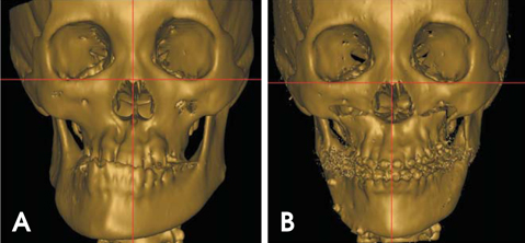
Fig. 1 CT scans from a 19-year-old female with class III malocclusion and facial asymmetry. A. Before orthognathic surgery. B. After orthognathic surgery. The menton deviation was reduced from 12.72 mm before surgery to 0.83 mm after surgery.
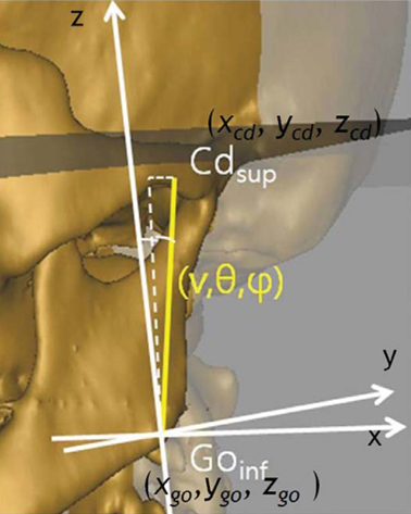
Fig. 2 A. Spherical coordinates of the analyzed facial lines and ramal height is shown. Length is indicated as v, the midsagittal inclination angle (between the line and the midsagittal reference plane) as θ, and the coronal inclination angle (between the line and the coronal reference plane) as φ.

Fig. 3 CT images of the facial lines before (A-H) and after (I-P) orthognathic surgery. A-D. The facial lines, ramal height and ramal lateral, before surgery. E-H. The facial lines, ramal posterior and mandibular body, before surgery. I-L. The facial lines, ramal height and ramal lateral, after surgery. M-P. The facial lines, ramal posterior and mandibular body, after surgery. The face remained three-dimensionally asymmetrical after surgery.
Reference
-
1. Aaronson SA. A cephalometric investigation of the surgical correction of mandibular prognathism. Angle Orthod. 1967; 37:251–260.2. Fromm B, Lundberg M. The soft-tissue facial profile before and after surgical correction of mandibular protrusion. Acta Odontol Scand. 1970; 28:157–177.
Article3. Robinson SW, Speidel TM, Isaacson RJ, Worms FW. Soft tissue profile change produced by reduction of mandibular prognathism. Angle Orthod. 1972; 42:227–235.4. Hershey HG, Smith LH. Soft-tissue profile change associated with surgical correction of the prognathic mandible. Am J Orthod. 1974; 65:483–502.
Article5. Fanibunda KB. Changes in the facial profile following correction for mandibular prognathism. Br J Oral Maxillofac Surg. 1989; 27:277–286.
Article6. Gjørup H, Athanasiou AE. Soft-tissue and dentoskeletal profile changes associated with mandibular setback osteotomy. Am J Orthod Dentofacial Orthop. 1991; 100:312–323.
Article7. McCance AM, Moss JP, Fright WR, James DR, Linney AD. A three dimensional analysis of soft and hard tissue changes following bimaxillary orthognathic surgery in skeletal III patients. Br J Oral Maxillofac Surg. 1992; 30:305–312.
Article8. Cho EJ, Yang WS. Soft tissue changes after double jaw surgery in skeletal class III malocclusion. Korean J Orthod. 1996; 26:1–16.9. Lin SS, Kerr WJ. Soft and hard tissue changes in Class III patients treated by bimaxillary surgery. Eur J Orthod. 1998; 20:25–33.
Article10. Soncul M, Bamber MA. Evaluation of facial soft tissue changes with optical surface scan after surgical correction of Class III deformities. J Oral Maxillofac Surg. 2004; 62:1331–1340.
Article11. Chew MT. Soft and hard tissue changes after bimaxillary surgery in Chinese Class III patients. Angle Orthod. 2005; 75:959–963.12. Park JW, Kim NK, Kim MJ, Chang YI. Three-dimensional analysis of soft and hard tissue changes after mandibular setback surgery in skeletal Class III patients. Korean J Orthod. 2005; 35:320–329.13. Sforza C, Peretta R, Grandi G, Ferronato G, Ferrario VF. Three-dimensional facial morphometry in skeletal Class III patients. A non-invasive study of soft-tissue changes before and after orthognathic surgery. Br J Oral Maxillofac Surg. 2007; 45:138–144.14. Baek SH, Cho IS, Chang YI, Kim MJ. Skeletodental factors affecting chin point deviation in female patients with class III malocclusion and facial asymmetry: a three-dimensional analysis using computed tomography. Oral Surg Oral Med Oral Pathol Oral Radiol Endod. 2007; 104:628–639.
Article15. Jung YJ, Kim MJ, Baek SH. Hard and soft tissue changes after correction of mandibular prognathism and facial asymmetry by mandibular setback surgery: three-dimensional using computerized tomography. Oral Surg Oral Med Oral Pathol Oral Radiol Endod. 2009; 107:763–771.16. Hwang HS, Hwang CH, Lee KH, Kang BC. Maxillofacial 3-dimensional image analysis for the diagnosis of facial asymmetry. Am J Orthod Dentofacial Orthop. 2006; 130:779–785.
Article17. Kim EJ, Palomo JM, Kim SS, Lim HJ, Lee KM, Hwang HS. Maxillofacial characteristics affecting chin deviation between mandibular retrusion and prognathism patients. Angle Orthod. 2011; 81:988–993.
Article18. Fisher NI, Lewis T, Embleton BJ. Statistical analysis of spherical data. Cambridge: Cambridge University Press;1987. p. 17–28.19. Yoon SJ, Wang RF, Hwang HS, Kang BC, Lee JS, Palomo JM. Application of spherical coordinate system to facial asymmetry analysis in mandibular prognathism patients. Imaging Sci Dent. 2011; 41:95–100.
Article20. Yoon SJ, Wang RF, Na HJ, Palomo JM. Normal range of facial asymmetry in spherical coordinates: a CBCT study. Imaging Sci Dent. 2013; 43:31–36.
Article21. Masuoka N, Muramatsu A, Ariji Y, Nawa H, Goto S, Ariji E. Discriminative thresholds of cephalometric indexes in the subjective evaluation of facial asymmetry. Am J Orthod Dentofacial Orthop. 2007; 131:609–613.
Article22. Haraguchi S, Takada K, Yasuda Y. Facial asymmetry in subjects with skeletal Class III deformity. Angle Orthod. 2002; 72:28–35.
- Full Text Links
-
- Actions
-
Cited
- CITED
-
- Close
- Share
-
- Similar articles
-
- Normal range of facial asymmetry in spherical coordinates: a CBCT study
- Quantification of three-dimensional facial asymmetry for diagnosis and postoperative evaluation of orthognathic surgery
- Unilateral intraoral vertical ramus osteotomy based on preoperative three-dimensional simulation surgery in a patient with facial asymmetry
- Practical utility of the three-dimensional approach in orthognathic surgery
- Current trends in orthognathic surgery
