Mandibular lateral incisor with four root canals: A unique case of double tooth diagnosed using multidetector computed tomography
- Affiliations
-
- 1Department of Conservative Dentistry and Endodontics, Manipal College of Dental Sciences, Manipal University, Manipal, India.
- 2Department of Conservative Dentistry and Endodontics, Faculty of Dentistry, Melaka Manipal Medical College, Manipal University, Manipal, India. drjayasiotia@yahoo.co.in
- KMID: 1974440
- DOI: http://doi.org/10.5624/isd.2013.43.2.123
Abstract
- Double tooth is a dental anomaly consequent to fusion of two or more teeth or gemination of a single tooth. This report describes a unique case of double tooth in relation to a mandibular lateral incisor exhibiting the presence of four root canals. The role of conventional radiography and advanced three-dimensional imaging techniques in the better assessment of complex root canal systems and their aid in endodontic management has also been highlighted.
MeSH Terms
Figure
-
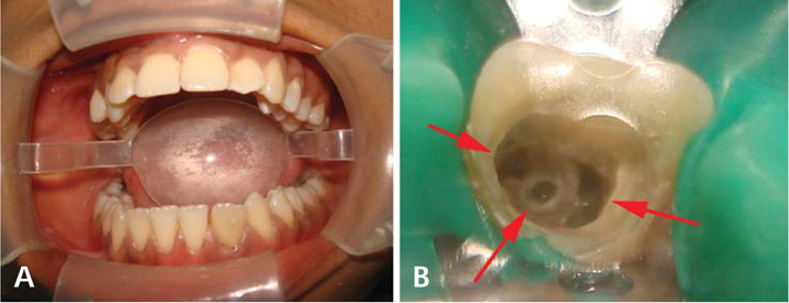
Fig. 1 A. Intraoral photograph shows a discolored mandibular left lateral incisor with an increased mesio-distal width. B. Three separate orifices of mesial, middle, and distal canals are seen in the access cavity (arrows).

Fig. 2 A. Preoperative intraoral periapical radiograph of the left mandibular lateral incisor shows multiple root canals in a single wide root with diffuse periapical radiolucency. B. The radiograph for working length determination. C. Post-obturation radiograph shows obturation of the distolingual, distobuccal, mesial, and middle canals (arrows). D. 1-year follow up radiograph shows satisfactory healing of the periapical lesion.

Fig. 3 A. Three-dimensional reconstruction of mandibular left lateral incisor (labial surface) shows a wide root. B. Three-dimensional reconstruction of the left mandibular lateral incisor shows a wide root with a lingual radicular groove (arrow).

Fig. 4 A. Axial CT image shows three canals at the cervical level: large distal, mesial, and middle canals. B. Axial image at the middle third of the root revealing the presence of two distal canals (arrows) and a single mesial canal. C. Axial image shows a narrow isthmus joining the mesial and distal canals.

Fig. 5 A. Curved coronal CT image shows a large distal and a mesial canal. B. Curved coronal image reveals three distinct root canals. C. Curved coronal image in which the connection between the root canals is evident.

Fig. 6 Sagittal CT images (A, B, and C) show the presence of aberrant root canal morphology and the presence of a large periapical radiolucency.
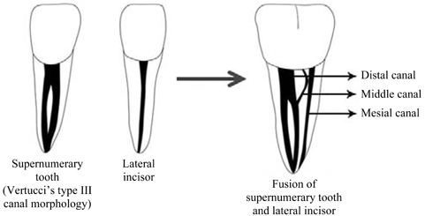
Fig. 7 Diagrammatic representation of the complex root canal morphology, as seen in the present case.
Reference
-
1. Duncan WK, Helpin ML. Bilateral fusion and gemination: a literature analysis and case report. Oral Surg Oral Med Oral Pathol. 1987. 64:82–87.
Article2. Brook AH, Winter GB. Double teeth. A retrospective study of 'geminated' and 'fused' teeth in children. Br Dent J. 1970. 129:123–130.
Article3. Saap JP, Eversole LR, Wysocki GP. Contemporary oral and maxillofacial pathology. 1997. St. Louis: Mosby.4. Chaudhry SI, Sprawson NJ, Howe L, Nairn RI. Dental twinning. Br Dent J. 1997. 182:185–188.
Article5. Tannenbaum KA, Alling EE. Anomalous tooth development. Case reports of gemination and twinning. Oral Surg Oral Med Oral Pathol. 1963. 16:883–887.6. Blaney TD, Hartwell GR, Bellizzi R. Endodontic management of a fused tooth: a case report. J Endod. 1982. 8:227–230.
Article7. Hülsmann M, Bahr R, Grohmann U. Hemisection and vital treatment of a fused tooth - literature review and case report. Endod Dent Traumatol. 1997. 13:253–258.8. Nunes E, de Moraes IG, de Novaes PM, de Sousa SM. Bilateral fusion of mandibular second molars with supernumerary teeth: case report. Braz Dent J. 2002. 13:137–141.
Article9. Schuurs AH, van Loveren C. Double teeth: review of the literature. ASDC J Dent Child. 2000. 67:313–325.10. Neville BW, Damm DD, Allen CM, Bouquet JE. Oral and maxillofacial pathology. 2009. 3rd ed. Philadelphia: WB Saunders;54–119.11. Peretz B, Brezniak N. Fusion of primary mandibular teeth: report of case. ASDC J Dent Child. 1992. 59:366–368.12. Camm JH, Wood AJ. Gemination, fusion and supernumerary tooth in the primary dentition: report of case. ASDC J Dent Child. 1989. 56:60–61.13. O'Reilly PM. Structural and radiographic evaluation of four cases of tooth fusion. Aust Dent J. 1990. 35:226–229.14. Itkin AB, Barr GS. Comprehensive management of the double tooth: report of case. J Am Dent Assoc. 1975. 90:1269–1272.
Article15. Pereira AJ, Fidel RA, Fidel SR. Maxillary lateral incisor with two root canals: fusion, gemination or dens invaginatus? Braz Dent J. 2000. 11:141–146.16. Onçag O, Candan U, Arikan F. Comprehensive therapy of a fusion between a mandibular lateral incisor and supernumerary tooth: case report. Int Dent J. 2005. 55:213–216.17. Peyrano A, Zmener O. Endodontic management of mandibular lateral incisor fused with supernumerary tooth. Endod Dent Traumatol. 1995. 11:196–198.
Article18. Mader CL. Fusion of teeth. J Am Dent Assoc. 1979. 98:62–64.
Article19. Salcido-García JF, Ledesma-Montes C, Hernández-Flores F, Pérez D, Garcés-Ortíz M. Frequency of supernumerary teeth in Mexican population. Med Oral Patol Oral Cir Bucal. 2004. 9:407–409.20. Lyroudia K, Mikrogeorgis G, Nikopoulos N, Samakovitis G, Molyvdas I, Pitas I. Computerized 3-D reconstruction of two "double teeth". Endod Dent Traumatol. 1997. 13:218–222.
Article21. Tsesis I, Steinbock N, Rosenberg E, Kaufman AY. Endodontic treatment of developmental anomalies in posterior teeth: treatment of geminated/fused teeth - report of two cases. Int Endod J. 2003. 36:372–379.
Article
- Full Text Links
-
- Actions
-
Cited
- CITED
-
- Close
- Share
-
- Similar articles
-
- Endodontic treatment of maxillary lateral incisors with anatomical variations
- Accuracy verification of dental cone-beam computed tomography of mandibular incisor root canals and assessment of its morphology and aging-related changes
- Nutrient canals on mandibular anterior region in cone beam computed tomography
- Unique case of a geminated supernumerary tooth with trifid crown
- Dilemmas pertaining to three canals in the mesiobuccal root of a maxillary second molar: a case report
