Endodontic treatment of maxillary lateral incisors with anatomical variations
- Affiliations
-
- 1Department of Conservative Dentistry, Kyungpook National University School of Dentistry, Daegu, Korea. skykim@knu.ac.kr
Abstract
- Maxillary lateral incisors usually exhibit a single root with a single canal. However, maxillary lateral incisor teeth with unusual morphology of root canal system are frequently reported. These cases of variable root canal anatomy can be treated well by nonsurgical endodontic methods. A detailed description of root canal morphology is fundamental for successful endodontic treatment. Treatment using an operating microscope, radiographs from different angles, and cone-beam computerized tomography (CBCT) can produce more predictable endodontic outcomes.
Figure
-
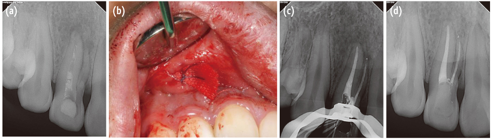
Figure 1 Right maxillary lateral incisor with two roots and two canals. (a) Preoperative periapical radiograph; (b) Intraoral photograph during incision and drainage; (c) Post-obturation periapical radiograph; (d) Periapical radiograph at the 6-month follow-up.

Figure 2 Left maxillary lateral incisor with one root and two canals. (a) Preoperative periapical radiograph; (b) Intraoral photograph of access cavity and canal orifices; (c) Post-obturation periapical radiograph; (d) Periapical radiograph at the 6-month follow-up.
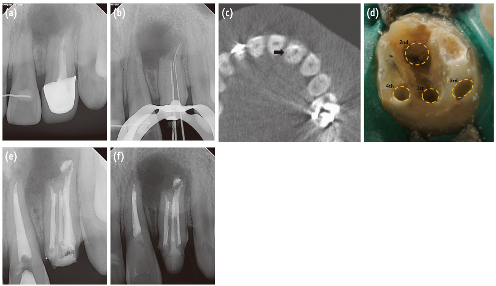
Figure 3 Left maxillary lateral incisor with one root and four canals. (a) Preoperative periapical radiograph; (b) Working length files of the first, second and third canals of the left maxillary lateral incisor; (c) Axial CBCT section of the tooth. Black arrow indicates the fourth canal; (d) Intraoral photograph of access cavity and canal orifices; (e) Postobturation periapical radiograph; (f) Periapical radiograph at the 4-month follow-up.
Reference
-
1. Vertucci FJ. Root canal anatomy of the human permanent teeth. Oral Surg Oral Med Oral Pathol. 1984; 58:589–599.
Article2. Walvekar SV, Behbehani JM. Three root canals and dens formation in a maxillary lateral incisor: a case report. J Endod. 1997; 23:185–186.
Article3. Pereira AJ, Fidel RA, Fidel SR. Maxillary lateral incisor with two root canals: fusion, germination or dens invaginatus? Braz Dent J. 2000; 11:141–146.4. Mohan AG, Rajesh EA, George L, Sujathan , Josy SA. Maxillary lateral incisors with two canals and two separate curved roots. Contemp Clin Dent. 2012; 3:519–521.
Article5. Lim YJ, Nam SH, Jung SH, Shin DR, Shin SJ, Min KS. Endodontic management of a maxillary lateral incisor with dens invaginatus and external root irregularity using cone-beam computed tomography. Restor Dent Endod. 2012; 37:50–53.
Article6. Calişkan MK, Pehlivan Y, Sepetçioğlu F, Türkün M, Tuncer SS. Root canal morphology of human permanent teeth in a Turkish population. J Endod. 1995; 21:200–204.
Article7. Sert S, Bayirli GS. Evaluation of the root canal configurations of the mandibular and maxillary permanent teeth by gender in the Turkish population. J Endod. 2004; 30:391–398.
Article8. Pineda F, Kuttler Y. Mesiodistal and buccolingual roentgenographic investigation of 7,275 root canals. Oral Surg Oral Med Oral Pathol. 1972; 33:101–110.
Article9. Kottoor J, Murugesan R, Albuquerque DV. A maxillary lateral incisor with four root canals. Int Endod J. 2012; 45:393–397.
Article10. Peix-Sánchez M, Miñana-Laliga R. A case of unusual anatomy: a maxillary lateral incisor with three canals. Int Endod J. 1999; 32:236–240.
Article11. Wei X, Senders C, Owiti GO, Liu X, Wei ZN, Dillard-Telm L, McClure HM, Hendrickx AG. The original and development of the upper lateral incisor and premaxilla in normal and cleft lip/palate monkeys induced with cyclophosphamide. Cleft Palate Craniofac J. 2000; 37:571–583.
Article12. Ferenczy K. The relationship of globulomaxillary cysts to the fusion of embryonal processes and to cleft palates. Oral Surg Oral Med Oral Pathol. 1958; 11:1388–1393.
Article13. Ooe T. On the early development of human dental lamina. Okajimas Folia Anat Jpn. 1957; 30:198–210.14. Lisson JA, Kjaer I. Location of alveolar clefts relative to the incisive fissure. Cleft Palate Craniofac J. 1997; 34:292–296.
Article15. Choi MS, Park SH, Cho KM, Kim JW. Treatment of a lateral incisor anatomically complicated with palatogingival groove. J Korean Acad Conserv Dent. 2011; 36:238–242.
Article16. Kim HI, Noh YS, Chang HS, Ryu HW, Min KS. The palato-gingival groove - anatomical anomaly occurred in maxillary lateral incisors: case reports. J Korean Acad Conserv Dent. 2007; 32:483–490.
Article17. Patel S, Dawood A, Ford TP, Whaites E. The potential applications of cone beam computed tomography in the management of endodontic problems. Int Endod J. 2007; 40:818–830.
Article18. Kim Y, Lee SJ, Woo J. Morphology of maxillary first and second molars analyzed by cone-beam computed tomography in a Korean population: variations in the number of roots and canals and the incidence of fusion. J Endod. 2012; 38:1063–1068.
Article
- Full Text Links
-
- Actions
-
Cited
- CITED
-
- Close
- Share
-
- Similar articles
-
- Surgical endodontic management of infected lateral canals of maxillary incisors
- A maxillary canine with two separated root canals: a case report
- The palato-gingival groove - anatomical anomaly occurred in maxillary lateral incisors: case reports
- Color Distribution of Maxillary Permanent Incisors in Korean Pediatric Patients Using a Spectrophotometer
- Color Comparison of Maxillary Primary Anterior Teeth and Various Composite Resins using a Spectrophotometer
