Unilateral segmental odontomaxillary hypoplasia: an unusual case report
- Affiliations
-
- 1Department of Oral Medicine and Radiology, Manipal College of Dental Sciences, Manipal, India. pandeysusma@gmail.com
- KMID: 1974399
- DOI: http://doi.org/10.5624/isd.2011.41.1.39
Abstract
- Facial asymmetry is not an uncommon occurrence in day to day dental practice. It can be caused by various etiologic factors ranging from facial trauma to serious hereditary conditions. Here, we report a rare case of non-syndromic facial asymmetry in a young female, who was born with this condition but was not aware of the progression of asymmetry. No relevant family history was recognized. She was also deficient in both deciduous and permanent teeth in the corresponding region of maxilla. Hence, the cause of this asymmetry was believed to be a segmental odontomaxillary hypoplasia of left maxilla accompanied by agenesis of left maxillary premolars and molars and disuse atrophy of corresponding facial musculature. This report briefly discussed the comparative features of segmental odontomaxillary hypoplasia, hemimaxillofacial dysplasia, and segmental odontomaxillary dysplasia and justified the differences between segmental odontomaxillary hypoplasia and the other two conditions.
Keyword
Figure
-
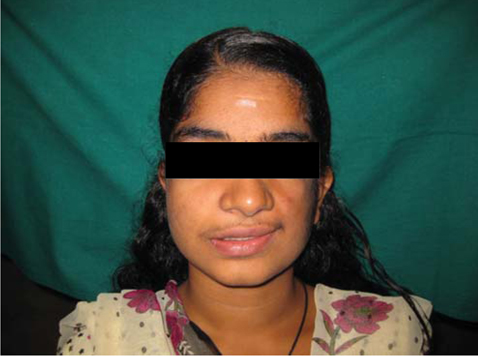
Fig. 1 Marked facial asymmetry with flattening and hypertrichosis of the middle third of left side of the face, incompetent lips, left angle of the mouth pulling upwards and mild drooping of the left eye.

Fig. 2 Over-retained left maxillary deciduous canine with clinically absence of all premolars and molars, and underdeveloped rudimentary corresponding alveolar ridge.
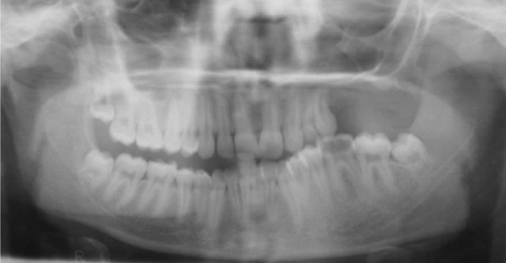
Fig. 3 Panoramic radiograph shows the absence of the left maxillary premolars and molars which associated with severe underdevelopment of the alveolar process. There is no missing tooth in the lower jaw. Lower left mandibular molars show supra eruption.

Fig. 4 Skull PA radiograph shows the maxillary deficiency in the middle third of the face on the left side. No other abnormalities of the lower jaw or any other facial bones were noted.
Reference
-
1. Proffit WR. Proffit WR, Fields HW, Sarver DM, editors. The etiology of orthodontic problems. Contemporary Orthodontics. 2006. 4th ed. St. Louis: C.V. Mosby;113–144.2. Prusack N, Pringle G, Scotti V, Chen SY. Segmental odontomaxillary dysplasia: a case report and review of the literature. Oral Surg Oral Med Oral Pathol Oral Radiol Endod. 2000. 90:483–488.3. Danforth RA, Melrose RJ, Abrams AM, Handlers JP. Segmental odontomaxillary dysplasia. Report of eight cases and comparison with hemimaxillofacial dysplasia. Oral Surg Oral Med Oral Pathol. 1990. 70:81–85.4. Becktor KB, Reibel J, Vedel B, Kjaer I. Segmental odontomaxillary dysplasia: clinical, radiological and histological aspect of four cases. Oral Dis. 2002. 8:106–110.5. Packota GV, Pharoah MJ, Petrikowski CG. Radiographic features of segmental odontomaxillary dysplasia: a study of 12 cases. Oral Surg Oral Med Oral Pathol Oral Radiol Endod. 1996. 82:577–584.6. Drake DL. Segmental odontomaxillary dysplasia: an unusual orthodontic challenge. Am J Orthod Dentofacial Orthop. 2003. 123:84–86.
Article
- Full Text Links
-
- Actions
-
Cited
- CITED
-
- Close
- Share
-
- Similar articles
-
- Two Cases of Unilateral Renal Hypoplasia
- Bilateral Segmental Neurofibromatosis with Partial Unilateral Lentiginosis: Case Report with Review of the Literature
- A Case of Curable Hypertension Owing to Unilateral Renal Hypoplasia
- Bilateral Segmental Vitiligo
- Isolated Unilateral Pulmonary Artery Hypoplasia with Accompanying Pulmonary Parenchymal Findings on CT: A Case Report
