Isolated Unilateral Pulmonary Artery Hypoplasia with Accompanying Pulmonary Parenchymal Findings on CT: A Case Report
- Affiliations
-
- 1Department of Radiology, Dongguk University Ilsan Hospital, Dongguk University College of Medicine, Goyang, Korea. sublime256@naver.com
- 2Department of Internal Medicine, Dongguk University Ilsan Hospital, Dongguk University College of Medicine, Goyang, Korea.
- 3Department of Radiology, Myongji Hospital, Goyang, Korea.
- KMID: 2377042
- DOI: http://doi.org/10.3348/jksr.2017.76.5.369
Abstract
- Unilateral pulmonary artery hypoplasia or agenesis without congenital cardiovascular anomalies is rare in adults. We report a case of a 36-year-old man with isolated left unilateral pulmonary artery hypoplasia with recurrent hemoptysis. On computed tomography (CT), the left pulmonary artery showed hypoplasia with multiple collateral vessels seen in the mediastinum and the left hemithorax. Also, parenchymal bands and peripheral linear opacities were seen in the affected lung, which were probably due to chronic infarction induced by unilateral pulmonary artery hypoplasia. There are only a few reports focusing on the radiologic findings in the pulmonary parenchyma induced by unilateral pulmonary artery hypoplasia, such as parenchymal bands and peripheral linear opacities. Therefore we report this case, which focused on the CT findings in the pulmonary parenchyma due to isolated unilateral pulmonary artery hypoplasia.
MeSH Terms
Figure
-
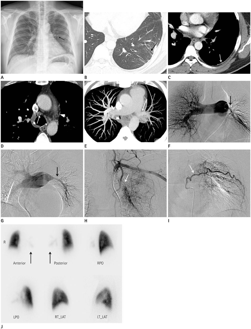
Fig. 1 Isolated unilateral pulmonary artery hypoplasia in a 36-year-old man who presented with hemoptysis. A. PA chest radiograph shows a small left hilum (arrow) compared to the right hilum. The left hemidiaphragm is slightly elevated, but mediastinal structures are not displaced. B-E. On chest CT with lung setting (B), parenchymal bands are seen in the posterolateral portion of the left lower lobe (arrow). Mediastinal setting of chest CT (C, D) shows serrated pleural thickening in the left posterolateral portion of the hemithorax (arrow). Tortuous prominent collateral vessels are noted in the mediastinum (arrows, arrowhead). Maximum intensity projection image (E) shows a hypoplastic left pulmonary artery (arrow). F-I. Pulmonary angiography (F, G) shows a small left pulmonary artery (arrows), compared to the right pulmonary artery. Left subclavian angiography (H) shows fine systemic collateral vessels (arrow) that supply the left lung. Left intercostal angiography (I) shows collateral branches of the left intercostal artery (arrow) with abnormal lung parenchymal staining. CT = computed tomography J. The lung perfusion scan shows a near total perfusion defect (arrows) in the left lung.
Cited by 1 articles
-
Spectrum of Multi-Detector Computed Tomography Findings that Alter Pulmonary Artery Diameters in Adults
Hye Rim Park, Young Tong Kim, Sung Shick Jou, Woong Hee Lee
J Korean Soc Radiol. 2018;78(6):389-403. doi: 10.3348/jksr.2018.78.6.389.
Reference
-
1. Chang Y, Ra SW, Chae EJ, Seo JB, Kim WY, Na S, et al. Isolated right pulmonary artery hypoplasia with retrograde blood flow in a 68-year old man. Tuberc Respir Dis. 2011; 71:126–133.2. Kadir IS, Thekudan J, Dheodar A, Jones MT, Carroll KB. Congenital unilateral pulmonary artery agenesis and aspergilloma. Ann Thorac Surg. 2002; 74:2169–2171.3. Cucci CE, Doyle EF, Lewis EW Jr. Absence of a primary division of the pulmonary trunk. An ontogenetic theory. Circulation. 1964; 29:124–131.4. Muthusami P, Ananthakrishnan R, Elangovan S. Incidentally detected unilateral pulmonary artery agenesis with pulmonary hypoplasia in a 67 year old woman. J Radiol Case Rep. 2010; 4:32–37.5. Shostak E, Sarwar A. A 50-year-old woman with dyspnea, lower extremity edema, and volume loss of the right hemithorax. Chest. 2009; 136:628–632.6. Sakai S, Murayama S, Soeda H, Furuya A, Ono M, Ro T, et al. Unilateral proximal interruption of the pulmonary artery in adults: CT findings in eight patients. J Comput Assist Tomogr. 2002; 26:777–783.7. Bouros D, Pare P, Panagou P, Tsintiris K, Siafakas N. The varied manifestation of pulmonary artery agenesis in adulthood. Chest. 1995; 108:670–676.8. Castañer E, Gallardo X, Ballesteros E, Andreu M, Pallardó Y, Mata JM, et al. CT diagnosis of chronic pulmonary thromboembolism. Radiographics. 2009; 29:31–50. discussion 50-53.9. Turner D, Vincent J, Epstein M. Isolated right pulmonary artery discontinuity. Images Paediatr Cardiol. 2000; 2:24–30.10. Reñé M, Sans J, Dominguez J, Sancho C, Valldeperas J. Unilateral pulmonary artery agenesis presenting with hemoptysis: treatment by embolization of systemic collaterals. Cardiovasc Intervent Radiol. 1995; 18:251–254.
- Full Text Links
-
- Actions
-
Cited
- CITED
-
- Close
- Share
-
- Similar articles
-
- Unilateral Absence of a Pulmonary Artery: Report of 3 cases
- Cystic Lung Changes in a Thin Section CT in an Asymptomatic Young Adult with Unilateral Pulmonary Vein Atresia: A Case Report
- A Case of Congenital Isolated Unilateral Absence of It. Pulmonary Artery
- Exogenous pulmonary surfactant replacement therapy in a neonate with pulmonary hypoplasia accompanying congenital diaphragmatic hernia: a case report
- A Case of Left Pulmonary Artery Hypoplasia in Adult
