Imaging Sci Dent.
2011 Mar;41(1):35-38. 10.5624/isd.2011.41.1.35.
Necrotizing sialometaplasia of palate: a case report
- Affiliations
-
- 1Department of Oral Medicine and Radiology, V.S Dental College and Hospital, Karnataka, India.
- 2Department of Oral Medicine, Diagnosis and Radiology, Dayanada Sagar College of Dental Sciences and Hospital, Karnataka, India. ramnarayanbk@gmail.com
- KMID: 1974398
- DOI: http://doi.org/10.5624/isd.2011.41.1.35
Abstract
- Necrotizing sialometaplasia (NS) which mimics malignancy both clinically and histopathologically is an uncommon benign, self-limiting inflammatory disease of the mucus-secreting minor salivary glands. The lesion is believed to be the result of vascular ischemia that may be initiated by trauma. Till date, the diagnosis of NS remains a challenge. This report demonstrates a case of NS in a 73-year-old male patient who presented with an ulcerative lesion in his palate. He had a history of local trauma and was long-term user of salbutamol inhaler. An incisional biopsy was carried out and the diagnosis was established through history, clinical examination, histopathology using Hematoxylin and Eosin stain. The patient was given symptomatic treatment and the lesion healed in about 7 weeks.
MeSH Terms
Figure
-
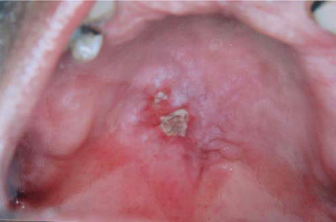
Fig. 1 The clinical photograph shows 2 irregular shaped ulcers in the posterior part of the palate with raised erythematous margins and surrounding mucosa appearing blanched, pale and grayish white. The underlying bone was partially exposed, and the floor was covered by yellowish grey slough.
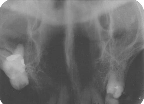
Fig. 2 Maxillary cross-sectional occlusal radiograph shows no bone involvement.
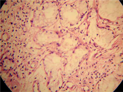
Fig. 3 Photomicrograph shows acinar necrosis under high magnification (H&E stain, 40×).
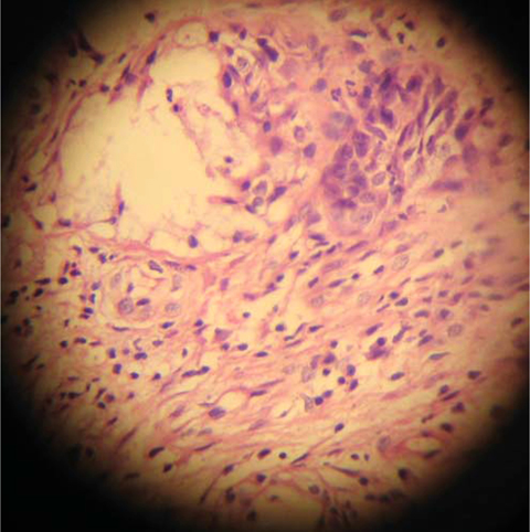
Fig. 4 Histopathological picture shows acinar necrosis and squamous metaplasia of salivary ductal cells (H&E stain, 40×).
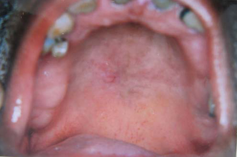
Fig. 5 Follow-up photograph after 6 weeks shows remission of the lesion except for an area of mild erythema.
Reference
-
1. Mesa ML, Gertler RS, Schneider LC. Necrotizing sialometaplasia: frequency of histologic misdiagnosis. Oral Surg Oral Med Oral Pathol. 1984. 57:71–73.
Article2. Abrams AM, Melrose RJ, Howell F. Necrotizing sialometaplasia. A disease simulating malignancy. Cancer. 1973. 32:130–135.
Article3. Brannon RB, Fowler CB, Hartman KS. Necrotizing sialometaplasia. A clinicopathologic study of sixty-nine cases and review of the literature. Oral Surg Oral Med Oral Pathol. 1991. 72:317–325.4. Schmidt-Westhausen A, Philipsen HP, Reichart PA. Necrotizing sialometaplasia of the palate. Literature report of 3 new cases. Dtsch Z Mund Kiefer Gesichtschir. 1991. 15:30–34.5. Randhawa T, Varghese I, Shameena PM, Sudha S, Nair RG. Necrotizing sialometaplasia of tongue. J Oral Maxillofac Pathol. 2009. 13:35–37.
Article6. Chen KT. Necrotizing sialometaplasia of the nasal cavity. Am J Otolaryngol. 1982. 3:444–446.
Article7. Walker GK, Fechner RE, Johns ME, Teja K. Necrotizing sialometaplasia of the larynx secondary to atheromatous embolization. Am J Clin Pathol. 1982. 77:221–223.
Article8. Schöning H, Emshoff R, Kreczy A. Necrotizing sialometaplasia in two patients with bulimia and chronic vomiting. Int J Oral Maxillofac Surg. 1998. 27:463–465.
Article9. Romagosa V, Bella MR, Truchero C, Moya J. Necrotizing sialometaplasia (adenometaplasia) of the trachea. Histopathology. 1992. 21:280–282.
Article10. Anneroth G, Hansen LS. Necrotizing sialometaplasia. The relationship of its pathogenesis to its clinical characteristics. Int J Oral Surg. 1982. 11:283–291.11. Zschoch H. Mucus gland infarct with squamous epithelial metaplasia in the lung. A rare site of so-called necrotizing sialometaplasia. Pathologe. 1992. 13:45–48.12. Hurt MA, Díaz-Arias AA, Rosenholtz MJ, Havey AD, Stephenson HE Jr. Posttraumatic lobular squamous metaplasia of breast. An unusual pseudocarcinomatous metaplasia resembling squamous (necrotizing) sialometaplasia of the salivary gland. Mod Pathol. 1988. 1:385–390.13. King DT, Barr RJ. Syringometaplasia: mucinous and squamous variants. J Cutan Pathol. 1979. 6:284–291.
Article14. Fowler CB, Brannon RB. Subacute necrotizing sialadenitis: report of 7 cases and a review of the literature. Oral Surg Oral Med Oral Pathol Oral Radiol Endod. 2000. 89:600–609.
Article15. Nah KS, Cho BH, Jung YH. Necrotizing sialometaplasia: report of 2 cases. Korean J Oral Maxillofac Radiol. 2006. 36:207–209.16. Daudia A, Murty GE. First case of full-thickness palatal necrotizing sialometaplasia. J Laryngol Otol. 2002. 116:219–220.
Article17. Keogh PV, O'Regan E, Toner M, Flint S. Necrotizing sialometaplasia: an unusual bilateral presentation associated with antecedent anaesthesia and lack of response to intralesional steroids. Case report and review of the literature. Br Dent J. 2004. 196:79–81.
Article18. Lamey PJ, Lewis MA, Crawford DJ, MacDonald DG. Necrotising sialometaplasia presenting as greater palatine nerve anaesthesia. Int J Oral Maxillofac Surg. 1989. 18:70–72.
Article19. Carlson DL. Necrotizing sialometaplasia: a practical approach to the diagnosis. Arch Pathol Lab Med. 2009. 133:692–698.
Article20. Gnepp DR. Warthin tumor exhibiting sebaceous differentiation and necrotizing sialometaplasia. Virchows Arch A Pathol Anat Histol. 1981. 391:267–273.
Article
- Full Text Links
-
- Actions
-
Cited
- CITED
-
- Close
- Share
-
- Similar articles
-
- Necrotizing Sialometaplasia Accompanied by Adenoid Cystic Carcinoma on the Soft Palate
- Necrotizing sialometaplasia: Report of 2 cases
- Contralateral recurrence of necrotizing sialometaplasia of the hard palate after five months: a case report
- A Case of Necrotizing Sialometaplasia
- A case of necrotizing sialomataplasia: consideration on cause, bone change, and incidence
