Ann Rehabil Med.
2011 Aug;35(4):499-506. 10.5535/arm.2011.35.4.499.
Foot Deformity in Charcot Marie Tooth Disease According to Disease Severity
- Affiliations
-
- 1Department and Research Institute of Rehabilitation Medicine, Yonsei University College of Medicine, Seoul 120-752, Korea. kimdy@yuhs.ac
- 2Department of Neurology, Ewha Woman's University School of Medicine, Seoul 158-710, Korea.
- KMID: 1971715
- DOI: http://doi.org/10.5535/arm.2011.35.4.499
Abstract
OBJECTIVE
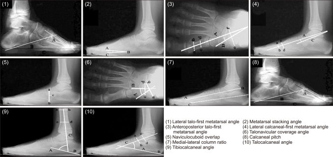
To investigate the characteristics of foot deformities in patients with Charcot-Marie-Tooth (CMT) disease compared with normal persons according to severity of disease. METHOD: Sixty-two patients with CMT disease were recruited for this study. The normal control group was composed of 28 healthy people without any foot deformity. Patients were classified into a mild group and a moderate group according to the CMT neuropathy score. Ten typical radiological angles representing foot deformities such as pes equinus and pes varus were measured. The CMT group angles were compared with those of the normal control group, and those of the mild group were also compared with those of the moderate group.
RESULTS
The lateral (Lat.) talo-first metatarsal angle, anteroposterior talo-first metatarsal angle, Lat. calcaneal-first metatarsal angle, Lat. naviocuboid overlap, Lat. calcaneal pitch, Lat. tibiocalcaneal angle, and Lat. talocalcaneal angle in the CMT group showed a significant difference compared to the normal control group (p<0.05). These findings revealed CMT patients have pes cavus, forefoot adduction, midfoot supination and pes varus deformity. Compared to the mild group, the moderate group significantly showed an increased Lat. calcaneal pitch and decreased Lat. calcaneal-first metatarsal angle, Lat. tibiocalcaneal angle, Lat. talocalcaneal angle, and Lat. talo-first metatarsal angle (p<0.05). These findings revealed that the pes cavus deformity of CMT patients tend to be worse with disease severity.
CONCLUSION
The characteristic equinovarus foot deformity patterns in CMT patients were revealed and these deformities tended to be worse with disease severity. Radiographic measures may be useful for the investigation of foot deformities in CMT patients.
Keyword
MeSH Terms
Figure
Reference
-
1. Dyck PJ, Lambert EH. Lower motor and primary sensory neuron diseases with peroneal muscular atrophy. I. Neurologic, genetic, and electrophysiologic findings in hereditary polyneuropathies. Arch Neurol. 1968; 18:603–618. PMID: 4297451.2. Madrid R, Bradley WG, Davis CJ. The peroneal muscular atrophy syndrome. Clinical, genetic, electrophysiological and nerve biopsy studies. Part 2. Observations on pathological changes in sural nerve biopsies. J Neurol Sci. 1977; 32:91–122. PMID: 864493.3. Apfel SC, Asbury AK, Bril V, Burns TM, Campbell JN, Chalk CH, Dyck PJ, Feldman EL, Fields HL, Grant IA, et al. Positive neuropathic sensory symptoms as endpoints in diabetic neuropathy trials. J Neurol Sci. 2001; 189:3–5. PMID: 11596565.4. Thomas PK, Calne DB. Motor nerve conduction velocity in peroneal muscular atrophy: evidence for genetic heterogeneity. J Neurol Neurosurg Psychiatry. 1974; 37:68–75. PMID: 4813428.
Article5. Buchthal F, Behse F. Peroneal muscular atrophy (PMA) and related disorders. I. Clinical manifestations as related to biopsy findings, nerve conduction and electromyography. Brain. 1977; 100:41–66. PMID: 861715.6. Nielsen VK, Pilgaard S. On the pathogenesis of Charcot-Marie-Tooth disease. A study of the sensory and motor conduction velocity in the median nerve. Acta Orthop Scand. 1972; 43:4–18. PMID: 5080641.7. Krajewski KM, Lewis RA, Fuerst DR, Turansky C, Hinderer SR, Garbern J, Kamholz J, Shy ME. Neurological dysfunction and axonal degeneration in Charcot-Marie-Tooth disease type 1A. Brain. 2000; 123:1516–1527. PMID: 10869062.
Article8. Sahenk Z, Chen L. Abnormalities in the axonal cytoskeleton induced by a connexin32 mutation in nerve xenografts. J Neurosci Res. 1998; 51:174–184. PMID: 9469571.
Article9. Skre H. Genetic and clinical aspects of Charcot-Marie-Tooth's disease. Clin Genet. 1974; 6:98–118. PMID: 4430158.
Article10. Holmes JR, Hansen ST Jr. Foot and ankle manifestations of Charcot-Marie-Tooth disease. Foot Ankle. 1993; 14:476–486. PMID: 8253442.
Article11. Jacobs JE, Carr CR. Progressive muscular atrophy of the peroneal type (Charcot-Marie-Tooth disease) orthopaedic management and end-result study. J Bone Joint Surg Am. 1950; 32A:27–38. PMID: 15401720.12. Mann DC, Hsu JD. Triple arthrodesis in the treatment of flxed cavovarus deformity in adolescent patients with Charcot-Marie-Tooth disease. Foot Ankle. 1992; 13:1–6. PMID: 1577335.
Article13. Aktas S, Sussman MD. The radiological analysis of pes cavus deformity in Charcot Marie Tooth disease. J Pediatr Orthop B. 2000; 9:137–140. PMID: 10868366.14. Chan G, Sampath J, Miller F, Riddle EC, Nagai MK, Kumar SJ. The role of the dynamic pedobarograph in assessing treatment of cavovarus feet in children with Charcot-Marie-Tooth disease. J Pediatr Orthop. 2007; 27:510–516. PMID: 17585258.
Article15. Shy ME, Rose MR. Charcot-Marie-Tooth disease impairs quality of life: Why? And how do we improve it? Neurology. 2005; 65:790–791. PMID: 16186514.
Article16. Gallardo E, Garcia A, Combarros O, Berciano J. Charcot-Marie-Tooth disease type 1A duplication: spectrum of clinical and magnetic resonance imaging features in leg and foot muscles. Brain. 2006; 129:426–437. PMID: 16317020.
Article17. Cornblath DR, Chaudhry V, Carter K, Lee D, Seysedadr M, Miernicki M, Joh T. Total neuropathy score: validation and reliability study. Neurology. 1999; 53:1660–1664. PMID: 10563609.
Article18. Azmaipairashvili Z, Riddle EC, Scavina M, Kumar SJ. Correction of cavovarus foot deformity in Charcot-Marie-Tooth disease. J Pediatr Orthop. 2005; 25:360–365. PMID: 15832156.
Article19. Sabir M, Lyttle D. Pathogenesis of pes cavus in Charcot-Marie-Tooth disease. Clin Orthop Relat Res. 1983; 175:173–178. PMID: 6839584.
Article20. Coleman SS, Chesnut WJ. A simple test for hindfoot flexibility in the cavovarus foot. Clin Orthop Relat Res. 1977; 123:60–62. PMID: 852192.
Article21. Mann RA, Missirain J. Pathophysiology of Charcot-Marie-Tooth disease. Clin Orthop Relat Res. 1988; 234:221–228. PMID: 3409580.
Article22. Tynan MC, Klenerman L, Helliwell TR, Edwards RH, Hayward M. Investigation of muscle imbalance in the leg in symptomatic forefoot pes cavus: a multidisciplinary study. Foot Ankle. 1992; 13:489–501. PMID: 1478577.
Article23. Swanson AB, Browne HS, Coleman JD. The cavus foot: concepts of production and treatment by metatarsal osteotomy. J Bone Joint Surg Am. 1966; 48:1019–1020.24. Hallgrimsson S. Pes cavus, seine behandlung und einige bemerkungen uber steinie atiologic. Acta Orthop Scand. 1939; 10:73–76.25. McCluskey WP, Lovell WW, Cummings RJ. The cavovarus foot deformity. Etiology and management. Clin Orthop Relat Res. 1989; 247:27–37. PMID: 2676298.26. Alexander IJ, Johnson KA. Assessment and management of pes cavus in Charcot-Marie-Tooth disease. Clin Orthop Relat Res. 1989; 246:273–281. PMID: 2766615.
Article27. Sala DA, Grant AD, Kummer FJ. Equinus deformity in cerebral palsy: recurrence after tendo Achillis lengthening. Dev Med Child Neurol. 1997; 39:45–48. PMID: 9003729.
Article
- Full Text Links
- Actions
-
Cited
- CITED
-
- Close
- Share
- Similar articles
-
- Operative Treatment of Charcot-Marie-Tooth Disease
- Misunderstanding of Foot Drop in a Patient with Charcot-Marie-Tooth Disease and Lumbar Disk Herniation
- A novel p.Leu699Pro mutation in MFN2 gene causes Charcot-Marie-Tooth disease type 2A
- Delayed Recovery of Neuromuscular Blockade by Rocuronium in a Patient with Charcot-Marie-Tooth Disease: Case reports
- A Case Report of Neuronal Type of Charcot-Marie-Tooth Disease


