Successful Treatment of Mycobacterium fortuitum Lung Disease with Oral Antibiotic Therapy: a Case Report
- Affiliations
-
- 1Division of Pulmonary and Critical Care Medicine, Department of Medicine, Samsung Medical Center, Sungkyunkwan University School of Medicine, Seoul, Korea. wjkoh@skku.edu
- KMID: 1970180
- DOI: http://doi.org/10.4046/trd.2008.64.4.293
Abstract
- Mycobacterium fortuitum usually causes colonization or transient infection in patients with underlying lung disease, such as prior tuberculosis or bronchiectasis. The majority of these patients may not need to receive antibiotic therapy for M. fortuitum isolates. We report here on a patient with M. fortuitum lung disease and who was successfully treated with combination oral antibiotic therapy. A 53-year-old woman was referred to our institution because of purulent sputum and dyspnea. A chest radiograph and computed tomography scan revealed cavitary consolidation in the left upper lobe and multiple small cavities in the left lower lobe. Numerous acid-fast bacilli (AFB) were seen in multiple sputum specimens and M. fortuitum was identified by culture from the sputum specimens. The patient received antibiotic treatment including clarithromycin, ciprofloxacin and sulfamethoxazole, because her symptoms were worsening despite conservative treatment. Sputum conversion was achieved after one month of antibiotic therapy. Both the patient's symptoms and radiographic findings improved after 10 months of antibiotic therapy.
MeSH Terms
Figure
-
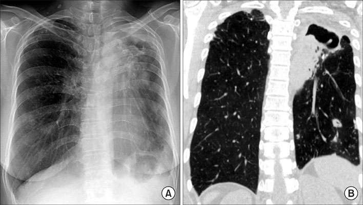
Figure 1 A 53-year-old woman with Mycobacterium fortuitum lung disease. The chest radiography shows a large cavitary consolidation in the left upper lobe (A). Reformatted coronal CT shows essentially the same findings as in A, with a large cavitary consolidation in the left upper lobe (B). Also note multiple nodules and consolidations in the left lower lobe.

Figure 2 A 53-year-old woman with Mycobacterium fortuitum lung disease. Before antibiotic treatment, chest CT shows the peribronchial consolidation in the lingular segment of the left upper lobe and multiple cavities in the left lower lobe (A, B). After 10 months of antibiotic treatment, chest CT shows improvement of the peribronchial consolidation (C, D). Also note improvement of multiple cavitary lesions in the left lower lobe.
Reference
-
1. Wolinsky E. Nontuberculous mycobacteria and associated diseases. Am Rev Respir Dis. 1979. 119:107–159.2. Brown TH. The rapidly growing mycobacteria-Mycobacterium fortuitum and Mycobacterium chelonei. Infect Control. 1985. 6:283–288.3. Wallace RJ Jr, Swenson JM, Silcox VA, Good RC, Tschen JA, Stone MS. Spectrum of disease due to rapidly growing mycobacteria. Rev Infect Dis. 1983. 5:657–679.4. Koh WJ, Kwon OJ, Jeon K, Kim TS, Lee KS, Park YK, et al. Clinical significance of nontuberculous mycobacteria isolated from respiratory specimens in Korea. Chest. 2006. 129:341–348.5. Awe RJ, Gangadharam PR, Jenkins DE. Clinical significance of Mycobacterium fortuitum infections in pulmonary disease. Am Rev Respir Dis. 1973. 108:1230–1234.6. Griffith DE, Girard WM, Wallace RJ Jr. Clinical features of pulmonary disease caused by rapidly growing mycobacteria: an analysis of 154 patients. Am Rev Respir Dis. 1993. 147:1271–1278.7. Park S, Suh GY, Chung MP, Kim H, Kwon OJ, Lee KS, et al. Clinical significance of Mycobacterium fortuitum isolated from respiratory specimens. Respir Med. 2008. 102:437–442.8. Lee SM, Kim J, Jeong J, Park YK, Bai GH, Lee EY, et al. Evaluation of the broth microdilution method using 2, 3-diphenyl-5-thienyl-(2)-tetrazolium chloride for rapidly growing mycobacteria susceptibility testing. J Korean Med Sci. 2007. 22:784–790.9. Daley CL, Griffith DE. Pulmonary disease caused by rapidly growing mycobacteria. Clin Chest Med. 2002. 23:623–632. vii10. Griffith DE, Aksamit T, Brown-Elliott BA, Catanzaro A, Daley C, Gordin F, et al. An official ATS/IDSA statement: diagnosis, treatment, and prevention of nontuberculous mycobacterial diseases. Am J Respir Crit Care Med. 2007. 175:367–416.11. Brown-Elliott BA, Wallace RJ Jr. Clinical and taxonomic status of pathogenic nonpigmented or late-pigmenting rapidly growing mycobacteria. Clin Microbiol Rev. 2002. 15:716–746.12. Vadakekalam J, Ward MJ. Mycobacterium fortuitum lung abscess treated with ciprofloxacin. Thorax. 1991. 46:737–738.13. Tasaka S, Urano T, Mori M, Yamaguchi K, Kanazawa M. A case of Mycobacterium fortuitum pulmonary disease in a healthy young woman successfully treated with ciprofloxacin and doxycycline. Kekkaku. 1995. 70:31–35.14. Ichiyama S, Tsukamura M. Ofloxacin and the treatment of pulmonary disease due to Mycobacterium fortuitum. Chest. 1987. 92:1110–1112.15. American Thoracic Society statement: diagnosis and treatment of disease caused by nontuberculous mycobacteria. Am J Respir Crit Care Med. 1997. 156:S1–S25.16. Kim SJ, Hong YP, Bai GH, Kim SC, Jin BW. Nontuberculous pulmonary infection in two patients with Mycobacterium avium-intracellulare complex and a patient with M. fortuitum. J Korean Soc Microbiol. 1982. 17:87–93.
- Full Text Links
-
- Actions
-
Cited
- CITED
-
- Close
- Share
-
- Similar articles
-
- A Case of Cutaneous Infection with Mycobacterium fortuitum
- Breast Abscess due to Mycobacterium Fortuitum: A Case Report
- Cutaneous Lesion due to Mycobacterium Fortuitum
- Spondylitis with an Epidural Abscess due to Mycobacterium fortuitum: A Case Report
- Mycobacterium fortuitum Infection Associated with Facial Fat Grafting: Simultaneous Infection of Liposuction and Liposculpture Site
