Multi-Focal Lobular Carcinoma In Situ Arising in Benign Phyllodes Tumor: A Case Report
- Affiliations
-
- 1Department of Radiology, Inha University Hospital, Incheon, Korea. kimyj@inha.ac.kr
- 2Department of Pathology, Inha University Hospital, Incheon, Korea.
- KMID: 1964265
- DOI: http://doi.org/10.3348/jksr.2015.73.2.127
Abstract
- Coexistent breast malignancy arising in phyllodes tumor is extremely rare, and most of them are incidental reports after surgical excision. Coexistent malignancy in phyllodes tumor can vary from in-situ to invasive carcinoma. Lobular neoplasia is separated into atypical lobular hyperplasia and lobular carcinoma in situ (LCIS). LCIS is known to have a higher risk of developing invasive cancer. We reported imaging findings of multifocal LCIS arising in benign phyllodes tumor.
Figure
-
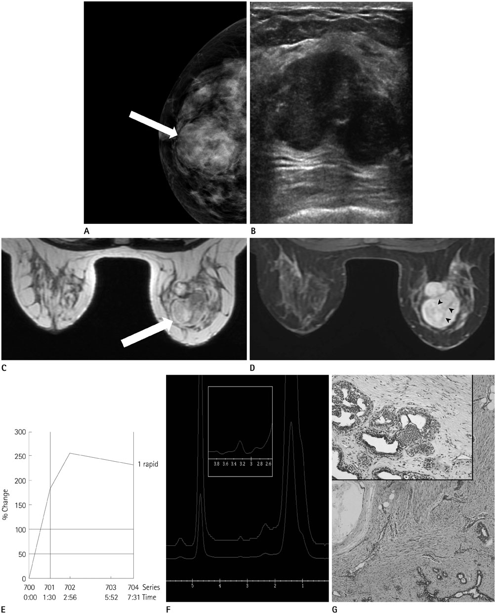
Fig. 1 A 41-year-old woman with multi-focal lobular carcinoma in situ in a phyllodes tumor. A. A craniocaudal mammogram shows a 4.16 × 3.70 × 3.80 cm sized, round mass with obscured margin and equal density in right subareolar breast at 12 o'clock (arrow). B. Breast ultrasonography shows relatively oval mass with heterogeneous echogenicity. C. T2-weighted axial MRI shows iso- and high-signal intense oval mass (arrow). D. Post-contrast T1-weighted MRI shows heterogeneous enhancement with dark non-enhancing internal septations (arrowheads) in the mass. E. Kinetics demonstrate initial fast (181%) followed by delayed washout (> 10%) with a fully automated computer-aided detection-based analysis. F. MR spectroscopy of the mass shows the presence of total choline containing compounds spectrum with a peak height at 3.2 ppm that was 2 fold above the baseline noise. G. The mass depicts 5 × 4-cm, benign phyllodes tumor with less than 3 mitotic figure/10 HPF and clear resection margins (H&E stain, × 40). Multi-focal lobular carcinoma in situ involves the stromal components of benign phyllodes tumor (inlet, H&E stain, × 200). H&E = hematoxylin and eosin
Reference
-
1. Shirah GR, Lau SK, Jayaram L, Bouton ME, Patel PN, Komenaka IK. Invasive lobular carcinoma and lobular carcinoma in situ in a phyllodes tumor. Breast J. 2011; 17:307–309.2. Padmanabhan V, Dahlstrom JE, Chong GC, Bennett G. Phyllodes tumor with lobular carcinoma in situ and liposarcomatous stroma. Pathology. 1997; 29:224–226.3. Nio Y, Iguchi C, Tsuboi K, Maruyama R. Ductal carcinoma in situ arising within a benign phyllodes tumor: a case report with a review of the literature. Oncol Lett. 2011; 2:223–228.4. Nomura M, Inoue Y, Fujita S, Sakao J, Hirota M, Souda S, et al. A case of noninvasive ductal carcinoma arising in malignant phyllodes tumor. Breast Cancer. 2006; 13:89–94.5. Shin DJ, Kim DB, Roh JH, Kwak BS. Ductal carcinoma in situ arising in a benign phyllodes tumor: a case report. J Korean Soc Radiol. 2013; 68:423–426.6. Deodhar KK, Baraniya JB, Naresh KN, Shinde SR, Chinoy RF. Cancerization of phyllodes tumour. Histopathology. 1997; 30:98–99.7. Tan H, Zhang S, Liu H, Peng W, Li R, Gu Y, et al. Imaging findings in phyllodes tumors of the breast. Eur J Radiol. 2012; 81:e62–e69.8. Kuhl CK, Mielcareck P, Klaschik S, Leutner C, Wardelmann E, Gieseke J, et al. Dynamic breast MR imaging: are signal intensity time course data useful for differential diagnosis of enhancing lesions? Radiology. 1999; 211:101–110.9. Tse GM, Yeung DK, King AD, Cheung HS, Yang WT. In vivo proton magnetic resonance spectroscopy of breast lesions: an update. Breast Cancer Res Treat. 2007; 104:249–255.
- Full Text Links
-
- Actions
-
Cited
- CITED
-
- Close
- Share
-
- Similar articles
-
- A Positive Hybrid (HMW-CK and E-Cadherin) Carcinoma in situ Arising in a Phyllodes Tumor of the Breast: A Case Report
- Borderline Phyllodes Tumor with an Incidental Invasive Tubular Carcinoma and Lobular Carcinoma In Situ Component: A Case Report
- Ductal Carcinoma In Situ Arising in a Benign Phyllodes Tumor: A Case Report
- Invasive Lobular Carcinoma of the Breast Associated with Mixed Lobular and Ductal Carcinoma In Situ: A Case Report
- Lobular carcinoma in situ in sclerosing adenosis
