Pial Arteriovenous Fistula with Giant Varices: Report of Two Cases with Good Surgical Outcome
- Affiliations
-
- 1Department of Neurosurgery, Shariati Hospital, Tehran University of Medical Sciences, Tehran, Iran. Sina21@gmail.com
- 2Department of Neurosurgery, Imam Khomeini Hospital, Tehran University of Medical Sciences, Tehran, Iran.
- KMID: 1963177
- DOI: http://doi.org/10.7461/jcen.2014.16.2.98
Abstract
- Pial arteriovenous fistulas (pAVF) are rare vascular lesions consisting of one or more arterial connections to a single venous channel without any intervening nidus of vessels or capillaries. Case 1: A 65-year-old woman with a complaint of headache and left hand paresthesia was referred to us. Magnetic resonance imaging showed a large saccular lesion with signal void in the posterior part of the right sylvian fissure and catheter angiography showed a giant venous aneurysm fed by one branch of the middle cerebral artery (MCA) and draining into the vein of Trolard. Case 2: A 12-year-old boy was transferred to our hospital with a history of sudden loss of consciousness and hemiplegia. Brain computed tomography revealed a massive hemorrhagic mass in the right hemisphere and cerebral angiography showed a pAVF with a large aneurysmal varix, which was fed by multiple branches of the right MCA and draining into the superior sagittal sinus. Both patients underwent craniotomy and after ligation of vascular connections, aneurysmal varices were removed completely. Surgical resection can be a safe method for treatment of pAVFs, particularly in those with large varices.
Keyword
MeSH Terms
Figure
-

Fig. 1 (A) Brain computed tomography showing a saccular mass with mural calcification in the right sylvian fissure. (B) Magnetic resonance imaging shows flow void within the lesion. (C) Right carotid angiogram (AP view) shows a giant venous aneurysm fed by one branch of the middle cerebral artery and draining via the vein of Trolard into the superior Sagittal sinus (black arrow). (D) After disconnection of the varix, it was resected totally. (E) Microscopic examination of the resected specimen reveals fragments of hyalinized and calcified vessles with myxoid changes.
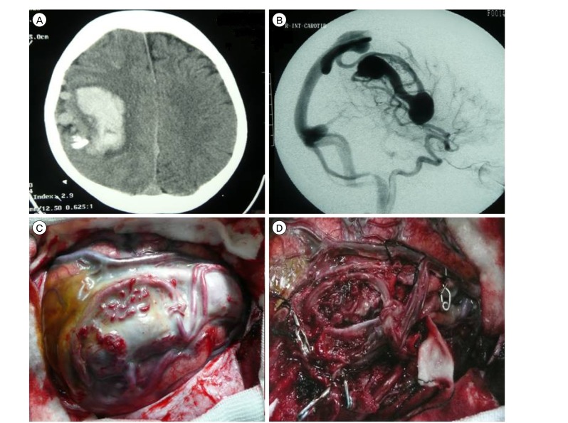
Fig. 2 (A) Brain computed tomography showing a hemorrhagic mass in the right parietal area with calcification and dilation of cortical vessels. (B) On lateral view of the right carotid angiogram, multiple branches of the middle cerebral artery are connected directly to the venous system and there is a large venous varix. (C) Intraoperative image shows the varix and associated vessels, which are surrounded by arachnoid. (D) After obliteration of feeding arteries (applied clips are evident) and draining vein, en bloc removal was achieved.
Reference
-
1. Almeida GM, Shibata MK. Hemispheric arteriovenous fistulae with giant venous dilation. Childs Nerv Syst. 1990; 6. 6(4):216–219. PMID: 2383876.
Article2. Aoki N, Sakai T, Oikawa A. Intracranial arteriovenous fistula manifesting as progressive neurological deterioration in an infant: case report. Neurosurgery. 1991; 4. 28(4):619–622. discussion 622-3. PMID: 2034363.
Article3. Barnwell SL, Ciricillo SF, Halbach VV, Edwards MS, Cogen PH. Intracerebral arteriovenous fistulas associated with intraparenchymal varix in childhood: case reports. Neurosurgery. 1990; 1. 26(1):122–125. PMID: 2294462.
Article4. Bendok BR, Getch CC, Frederiksen J, Batjer HH. Resection of a large arteriovenous fistula of the brain using low-flow deep hypothermic cardiopulmonary bypass: technical case report. Neurosurgery. 1999; 4. 44(4):888–890. discussion 890-1. PMID: 10201318.
Article5. Carrillo R, Carreira LM, Prada J, Rosas C, Egas G. Giant aneurysm arising from a single arteriovenous fistula in a child. Case report. J Neurosurg. 1984; 5. 60(5):1085–1088. PMID: 6716144.6. Garcia-Monaco R, de Victor D, Mann C, Hannedouche A, Terbrugge K, Lasjaunias P. Congestive cardiac manifestations from cerebrocranial arteriovenous shunts. Endovascular management in 30 children. Childs Nerv Syst. 1991; 2. 7(1):48–52. PMID: 2054809.7. Garcia-Monaco R, Taylor W, Rodesch G, Alvarez H, Burrows P, Coubes P, et al. Pial arteriovenous fistula in children as presenting manifestation of Rendu-Osler-Weber disease. Neuroradiology. 1995; 1. 37(1):60–64. PMID: 7708192.
Article8. Giller CA, Batjer HH, Purdy P, Walker B, Mathews D. Interdisciplinary evaluation of cerebral hemodynamics in the treatment of arteriovenous fistulae associated with giant varices. Neurosurgery. 1994; 10. 35(4):778–782. discussion 782-4. PMID: 7808630.
Article9. Halbach VV, Higashida RT, Hieshima GB, Hardin CW, Dowd CF, Barnwell SL. Transarterial occlusion of solitary intracerebral arteriovenous fistulas. AJNR Am J Neuroradiol. 1989; Jul-Aug. 10(4):747–752. PMID: 2505503.10. Hermier M, Turjman F, Bozio A, Duquesnel J, Lapras C. Endovascular treatment of an infantile nongalenic cerebral arteriovenous fistula with cyanoacrylate. Childs Nerv Syst. 1995; 8. 11(8):494–498. PMID: 7585691.
Article11. Hoh BL, Putman CM, Budzik RF, Ogilvy CS. Surgical and endovascular flow disconnection of intracranial pial single-channel arteriovenous fistulae. Neurosurgery. 2001; 12. 49(6):1351–1363. discussion 1363-4. PMID: 11846934.
Article12. Kikuchi K, Kowada M, Sasajima H. Vascular malformations of the brain in hereditary hemorrhagic telangiectasia (Rendu-Osler-Weber disease). Surg Neurol. 1994; 5. 41(5):374–380. PMID: 8009411.
Article13. Lee JS, Oh CW, Bang JS, Kwon OK, Hwang G. Intracranial pial arteriovenous fistula presenting with hemorrhage: a case report. J Cerebrovasc Endovasc Neurosurg. 2012; 12. 14(4):305–308. PMID: 23346547.
Article14. Lownie S, Duckwiler G, Fox A, Drake C. Endovascular therapy of nongalenic cerebral arteriovenous fistulas. In : Viñuela F, Halbach V, Dion J, editors. Interventional neuroradiology: endovascular therapy of the central nervous system. New York: Raven Press;1992. p. 87–106.15. Lv X, Li Y, Jiang C, Wu Z. Endovascular treatment of brain arteriovenous fistulas. AJNR Am J Neuroradiol. 2009; 4. 30(4):851–856. PMID: 19147710.
Article16. Meyer FB, Grady RE, Abel MD, Nichols DA, Caminha SS, Robb RA, et al. Resection of a large temporooccipital parenchymal arteriovenous fistula by using deep hypothermic circulatory bypass. Case report. J Neurosurg. 1997; 12. 87(6):934–939. PMID: 9384407.17. Morimoto T, Yamada T, Hashimoto H, Tokunaga H, Tsunoda S, Sakaki T. Direct approach to intracranial vertebral arteriovenous fistula. Case report. Acta Neurochir (Wien). 1995; 137(1-2):98–101. PMID: 8748878.18. Nelson K, Nimi Y, Lasjaunias P, Berenstein A. Endovascular embolization of congenital intracranial pial arteriovenous fistulas. Neuroimaging Clin N Am. 1992; 2:309–317.19. Oh HJ, Yoon SM, Kim SH, Shim JJ. A case of pial arteriovenous fistula with giant venous aneurysm and multiple varices treated with coil embolization. J Korean Neurosurg Soc. 2011; 9. 50(3):248–251. PMID: 22102958.
Article20. Passacantilli E, Pichierri A, Guidetti G, Santoro A, Delfini R. Surgical treatment of pial cerebellar arteriovenous fistulas with aneurysm of the main feeding artery. Surg Neurol. 2006; 1. 65(1):90–94. PMID: 16378872.
Article21. Ratliff J, Voorhies RM. Arteriovenous fistula with associated aneurysms coexisting with dural arteriovenous malformation of the anterior inferior falx. Case report and review of the literature. J Neurosurg. 1999; 8. 91(2):303–307. PMID: 10433319.22. Santosh C, Teasdale E, Molyneux A. Spontaneous closure of an intracranial middle cerebral arteriovenous fistula. Neuroradiology. 1991; 33(1):65–66. PMID: 2027449.
Article23. Tabatabai SA, Zadeh MZ, Habibi Z, Meybodi AT, Hashemi M. Intracerebral atypical calcification in nongalenic pial arteriovenous fistula: a case report. Cases J. 2008; 11. 1(1):335. PMID: 19019250.
Article24. Talamonti G, Versari PP, DAliberti G, Villa F, Fontana RA, Collice M. Complex arteriovenous fistula of the brain in an infant Case report. J Neurosurg Sci. 1997; 12. 41(4):337–341. PMID: 9555640.25. Tomlinson FH, Rufenacht DA, Sundt TM Jr, Nichols DA, Fode NC. Arteriovenous fistulas of the brain and the spinal cord. J Neurosurg. 1993; 7. 79(1):16–27. PMID: 8315463.
Article26. Vinuela F, Drake CG, Fox AJ, Pelz DM. Giant intracranial varices secondary to high-flow arteriovenous fistulae. J Neurosurg. 1987; 2. 66(2):198–203. PMID: 3806202.27. Yamashita K, Ohe N, Yoshimura S, Iwama T. Intracranial pial arteriovenous fistula. Neurol Med Chir (Tokyo). 2007; 12. 47(12):550–554. PMID: 18159139.28. Yang WH, Lu MS, Cheng YK, Wang TC. Pial arteriovenous fistula: a review of literature. Br J Neurosurg. 10. 25(5):580–585. PMID: 21501060.
Article29. Yasargil MG. Microneurosurgery. Stuttgart: Georg Thieme Verlag;1993.
- Full Text Links
-
- Actions
-
Cited
- CITED
-
- Close
- Share
-
- Similar articles
-
- Successful Treatment of Intracranial Small Pial Single-Channel Arteriovenous Fistula Using N-butyl Cyanoacrylate: Report of 2 Cases
- Embolization of Cerebral Pial Arteriovenous Fistula Under Balloon-assisted Flow Control Using NBCA: a Case Report
- A Case of Pial Arteriovenous Fistula with Giant Venous Aneurysm and Multiple Varices Treated with Coil Embolization
- Coil Embolization of High-flow Pial Arteriovenous Fistula and Management of Hyperperfusion Syndrome: a Case Report
- Congenital Intracranial Pial Arteriovenous Fistula Complicated with Congestive Heart Failure in Neonate: A Case Report
