An atypical case of rare salivary malignancy, hyalinizing clear cell carcinoma
- Affiliations
-
- 1Department of Oral and Maxillofacial Surgery, College of Dentistry, Yonsei University, Seoul, Korea. omsnam@yuhs.ac
- 2Oral Cancer Research Institute, College of Dentistry, Yonsei University, Seoul, Korea.
- 3Department of Oral Pathology, College of Dentistry, Yonsei University, Seoul, Korea.
- KMID: 1960978
- DOI: http://doi.org/10.5125/jkaoms.2013.39.6.283
Abstract
- As an uncommon, malignant salivary gland tumor with female predominance, hyalinizing clear cell carcinoma (HCCC) is regarded as an indolent tumor. The diagnosis of this rare tumor is challenging, and it depends on microscopic and immunohistochemical (IHC) studies. Although it is regarded as an indolent tumor, there are reports of unconventional forms with aggressive clinical courses. We report an atypical case of this rare tumor, HCCC, in a male patient who had a relatively large-sized mass (3.8x3.0 cm) on the right mouth floor with ipsilateral neck node metastasis. The clinical, radiological, pathological, and IHC features together with the clinical course are described.
MeSH Terms
Figure
-
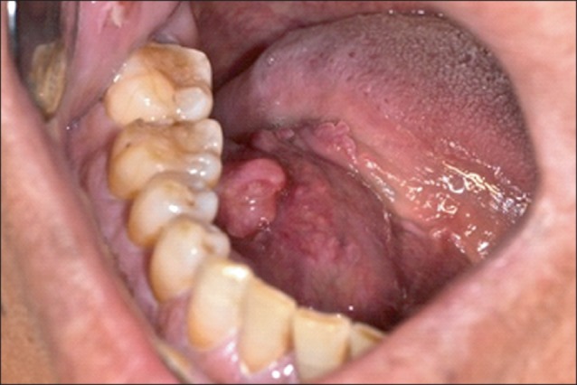
Fig. 1 Initial intraoral photograph.
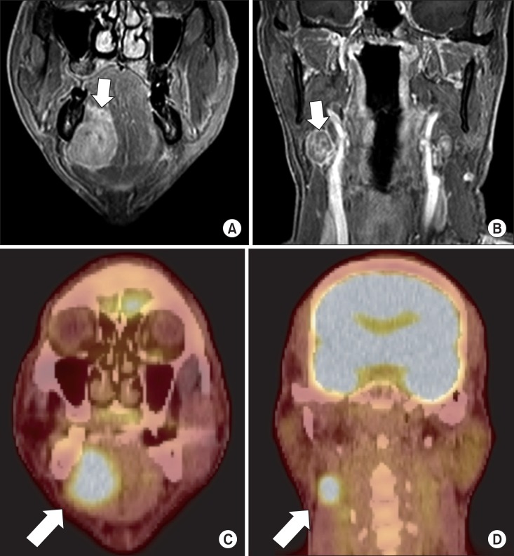
Fig. 2 Gadolinium-enhanced magnetic resonance imaging, coronal view. A. Main mass (arrow). B. Nodal metastasis on level II, ipsilateral side (arrow). C. Positron emission tomography-computed tomography, coronal view. Main mass (arrow). D. Nodal metastasis on level II, ipsilateral side (arrow).
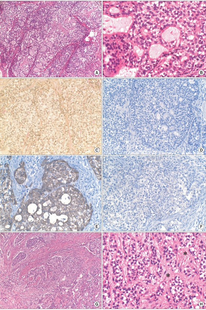
Fig. 3 A. Histopathological examination of incisional biopsy. Poorly differentiated epithelial cells with multiple clear cells were noted (H&E staining, ×100). B. Specimen from incisional biopsy in higher magnification. The specimen was mostly composed of malignant cells with clear cytoplasm, and there were some portions of cells with glandular differentiation (H&E staining, ×400). C. The specimen from incisional biopsy showing clear cells stained negatively to mucicarmine (×200). D. Immunohistochemical (IHC) staining for S-100 showing negative reactivity of clear cells (×200). E. IHC staining for cytokeratin (CK) 7 showing positive reactivity of clear cells (×200). F. IHC staining for CK20 showing negative reactivity of clear cells (×200). G. Histopathologic examination of final specimen. Epithelial cells and clear cells arranged in cords or nests (H&E staining, ×100). H. Final specimen in higher magnification. Areas of hyalinization are seen (marked in asterisks) (H&E staining, ×400).
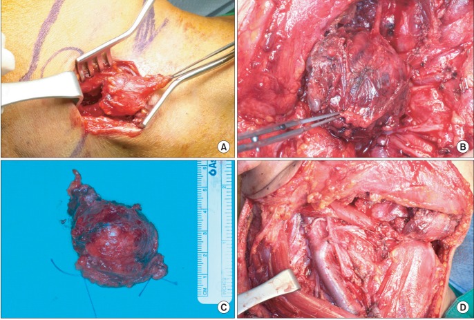
Fig. 4 Intraoperative photographs. A. Palpable node on level II was revealed. B. The main lesion was shown. C. Surgical specimen of the main mass. D. Operation site after the excision and neck dissection.
Reference
-
1. Dardick I, Leong I. Clear cell carcinoma: review of its histomorphogenesis and classification as a squamous cell lesion. Oral Surg Oral Med Oral Pathol Oral Radiol Endod. 2009; 108:399–405. PMID: 19570692.
Article2. Milchgrub S, Gnepp DR, Vuitch F, Delgado R, Albores-Saavedra J. Hyalinizing clear cell carcinoma of salivary gland. Am J Surg Pathol. 1994; 18:74–82. PMID: 7506496.
Article3. Baghirath PV, Kumar JV, Vinay BH. Hyalinizing clear cell carcinoma: a rare entity. J Oral Maxillofac Pathol. 2011; 15:335–339. PMID: 22144841.
Article4. O'Sullivan-Mejia ED, Massey HD, Faquin WC, Powers CN. Hyalinizing clear cell carcinoma: report of eight cases and a review of literature. Head Neck Pathol. 2009; 3:179–185. PMID: 20596970.5. O'Regan E, Shandilya M, Gnepp DR, Timon C, Toner M. Hyalinizing clear cell carcinoma of salivary gland: an aggressive variant. Oral Oncol. 2004; 40:348–352. PMID: 14747068.6. Savera AT, Sloman A, Huvos AG, Klimstra DS. Myoepithelial carcinoma of the salivary glands: a clinicopathologic study of 25 patients. Am J Surg Pathol. 2000; 24:761–774. PMID: 10843278.7. Meer S, Altini M. CK7+/CK20- immunoexpression profile is typical of salivary gland neoplasia. Histopathology. 2007; 51:26–32. PMID: 17593078.
Article8. Ellis GL. Clear cell neoplasmsin salivary glands: clearly a diagnostic challenge. Ann Diagn Pathol. 1998; 2:61–78. PMID: 9845723.9. Said-Al-Naief N, Klein MJ. Clear cell entities of the head and neck: a selective review of clear cell tumors of the salivary glands. Head Neck Pathol. 2008; 2:111–115. PMID: 20614333.
Article10. Batsakis JG, el-Naggar AK, Luna MA. Hyalinizing clear cell carcinoma of salivary origin. Ann Otol Rhinol Laryngol. 1994; 103:746–748. PMID: 8085740.
Article11. Tang SK, Wan SK, Chan JK. Hyalinizing clear cell carcinoma of salivary gland: report of a case with multiple recurrences over 12 years. Am J Surg Pathol. 1995; 19:240–241. PMID: 7530410.
- Full Text Links
-
- Actions
-
Cited
- CITED
-
- Close
- Share
-
- Similar articles
-
- Hyalinizing Clear Cell Carcinoma of the Base of Tongue Mistaken for Benign Lesion: A Case Report
- A Rare Case of Clear Cell Carcinoma of the Tongue Base: A Case Report
- A Case of Hyalinizing Trabecular Tumor of the Thyroid Gland
- Fine Needle Aspiration Cytology of Papillary-Cystic Variant of Acinic Cell Carcinoma of Salivary Gland: A Case Report
- A Case of Clear Cell Squamous Cell Carcinoma
