Unilateral Chest Wall Anomaly in a Patient with Gardner's Syndrome: Case Report
- Affiliations
-
- 1Department of Radiology, Kangbuk Samsung Hospital, Sungkyunkwan University School of Medicine, Seoul, Korea. capella27@gmail.com
- 2Department of Radiology, Eulji General Hospital, Eulji University School of Medicine, Seoul, Korea.
- KMID: 1941785
- DOI: http://doi.org/10.3348/jksr.2014.70.5.373
Abstract
- Gardner syndrome is a familial disease consisting of colonic polyposis, osteomas, and soft tissue tumors. We describe unilateral chest wall anomaly in a 32-year-old man with Gardner syndrome. A chest radiograph showed asymmetric hypertrophy of the right seventh to tenth ribs. CT images showed increased size of the medullary portions of these lesions, but relatively normal thickness of the cortex. Intercostal muscles along the right seventh to tenth ribs were hypertrophied as compared with the contralateral ribs. Both lungs were clear but the volume of right lung showed slightly smaller than left one.
MeSH Terms
Figure
-

Fig. 1 Colonoscopy and endoscopy in a 32-year-old man with Gardner's syndrome. A. Colonoscopy reveals extensive polypoid lesions along the entire colon. B. A 5 cm sized polypoid mass is noted 10 cm above the anal verge, which was confirmed as being a tubular adenoma with low grade dysplasia following surgical excision. C. Endoscopy shows numerous small polypoid lesions on the gastric wall, which are confirmed by endoscopic biopsy as being tubular adenomas with low grade dysplasia.
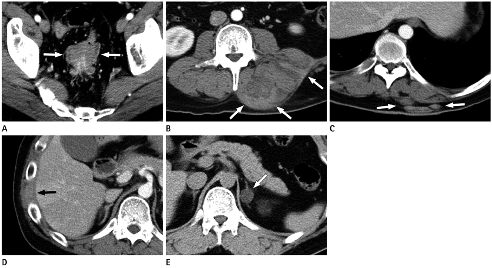
Fig. 2 Postcontrast CT of the abdomen in a 32-year-old man with Gardner's syndrome. A. Axial postcontrast CT scan shows an intraluminal polypoid mass (arrows) with perirectal infiltration in the upper rectum, which was confirmed as being a tubular adenoma with low grade dysplasia following surgical excision (same lesion with Fig. 1B). B-D. Axial postcontrast CT images, showing soft tissue tumors of heterogenous density (arrows) around the left paraspinal and quadratus lumborum muscles (B), the subcutaneous fat layer of left thorax (C), and along the right ninth intercostal muscle (D), suggesting desmoids tumors. E. Axial noncontrast CT scan, showing a left adrenal mass (arrow) of less than 10 Hounsfield units, suggesting an adrenal adenoma.
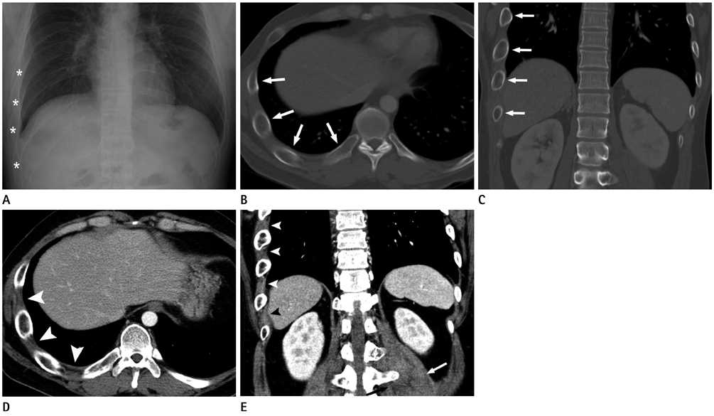
Fig. 3 CT and radiography of the chest in a 32-year-old man with Gardner's syndrome. A. Posteroanterior plain radiograph of the chest, showing unilateral hypertrophy of the right seventh through tenth ribs (asterisks). B, C. Axial (B) and coronal (C) CT images with bone window shows increased size of the medullary portion of the involved ribs (arrows) with relatively normal thickness of the cortex. D, E. Axial (D) and coronal (E) CT images with soft tissue window shows increased size of the intercostals muscles (arrowheads) as compared with contralateral side. Left paraspinal muscle shows swelling with ill-defined mass-like lesion (arrows) due to desmoids tumor (same lesion with Fig. 2B).
Reference
-
1. Rai AT, Nguyen TP, Hogg JP, Gabriele FJ. Aggressive fibromatosis of the neck in a patient with Gardner's syndrome. Neuroradiology. 2001; 43:650–652.2. Traill Z, Tuson J, Woodham C. Adrenal carcinoma in a patient with Gardner's syndrome: imaging findings. AJR Am J Roentgenol. 1995; 165:1460–1461.3. Hatzimarkou A, Filippou D, Papadopoulos V, Filippou G, Rizos S, Skandalakis P. Desmoid tumor in Gardner's Syndrome presented as acute abdomen. World J Surg Oncol. 2006; 4:18.4. Fotiadis C, Tsekouras DK, Antonakis P, Sfiniadakis J, Genetzakis M, Zografos GC. Gardner's syndrome: a case report and review of the literature. World J Gastroenterol. 2005; 11:5408–5411.5. Ishida T, Abe S, Miki Y, Imamura T. Intraosseous pilomatricoma: a possible rare skeletal manifestation of Gardner syndrome. Skeletal Radiol. 2007; 36:693–698.6. Chang CH, Piatt ED, Thomas KE, Watne AL. Bone abnormalities in Gardner's syndrome. Am J Roentgenol Radium Ther Nucl Med. 1968; 103:645–652.7. Harned RK, Buck JL, Olmsted WW, Moser RP, Ros PR. Extracolonic manifestations of the familial adenomatous polyposis syndromes. AJR Am J Roentgenol. 1991; 156:481–485.8. Dolan KD, Seibert J, Seibert RW. Gardner's syndrome. A model for correlative radiology. Am J Roentgenol Radium Ther Nucl Med. 1973; 119:359–364.9. Nandakumar G, Morgan JA, Silverberg D, Steinhagen RM. Familial polyposis coli: clinical manifestations, evaluation, management and treatment. Mt Sinai J Med. 2004; 71:384–391.
