Osteochondroma of the 5th and 6th Cervical Vertebral Body: One Case Report
- Affiliations
-
- 1Department of Orthopaedic Surgery, Our Lady of Mercy Hospital, College of Medicine, the Catholic University of Korea. drcst@lycos.co.kr
- KMID: 1941691
- DOI: http://doi.org/10.4184/jkss.2005.12.3.238
Abstract
- Here, the case of an osteochondroma, which developed on the cervical spine of an 18-year old boy, is presented. Generally, an osteochondroma is very difficult to diagnose, due to their rarity and non-specific or lack of symptoms, and because they show radiolucency on plain X-ray. Due to the neurological symptoms of this patient, including radiating pain and a palpable protruding hard mass, the CT and MRI images were checked for a more accurate radiological evaluation. These images showed evidence of spinal cord compression or obliteration of the neural foramen. An en bloc excisional biopsy of the bony mass and cartilage cap, and a decompressive laminectomy were performed.
Keyword
MeSH Terms
Figure
-
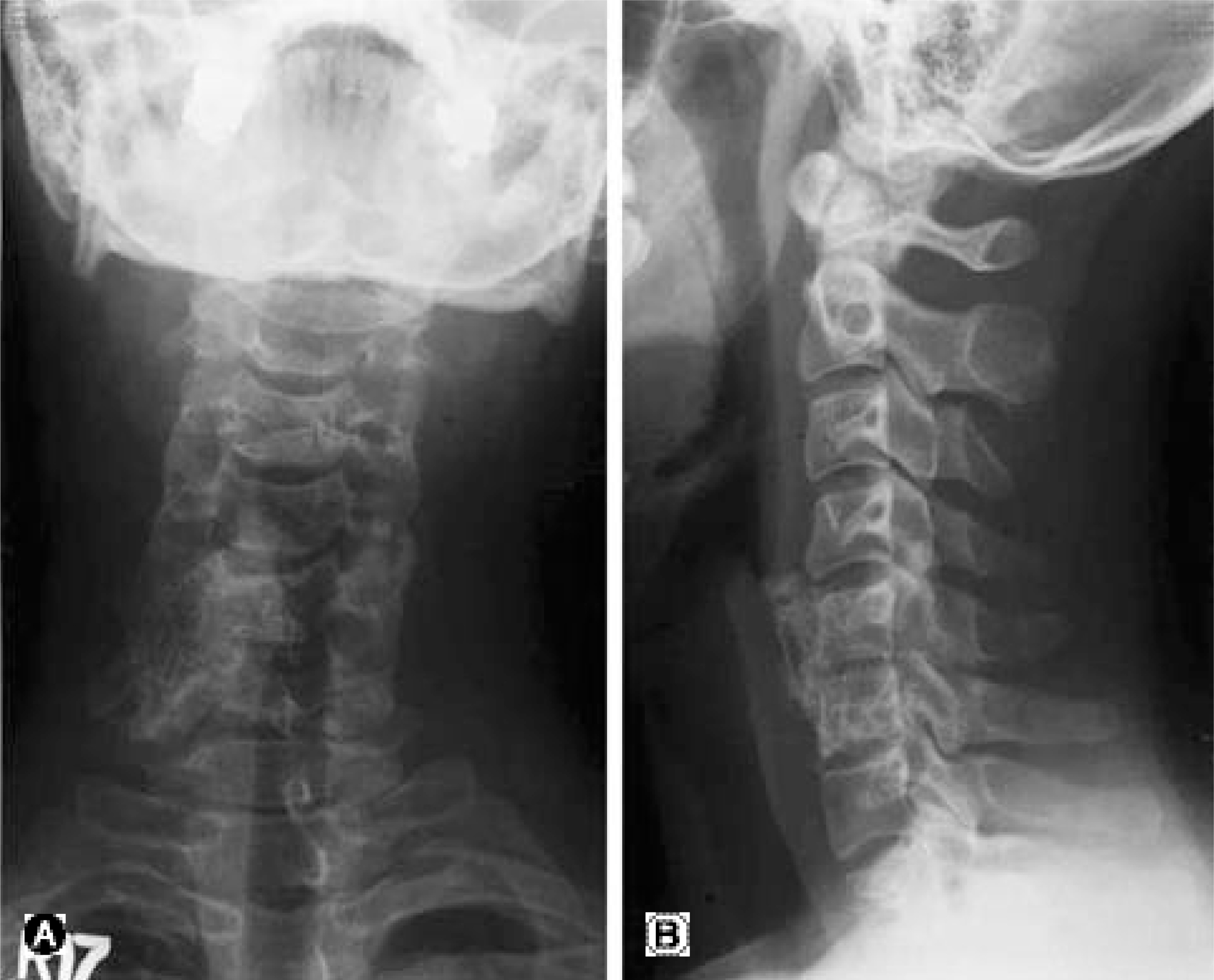
Fig. 1. It shows amorphous bony mass contained irregular bony trabeculae in right lateral portion of the 5-6 cervical spine on plain X-ray anteroposterior view and lateral view.
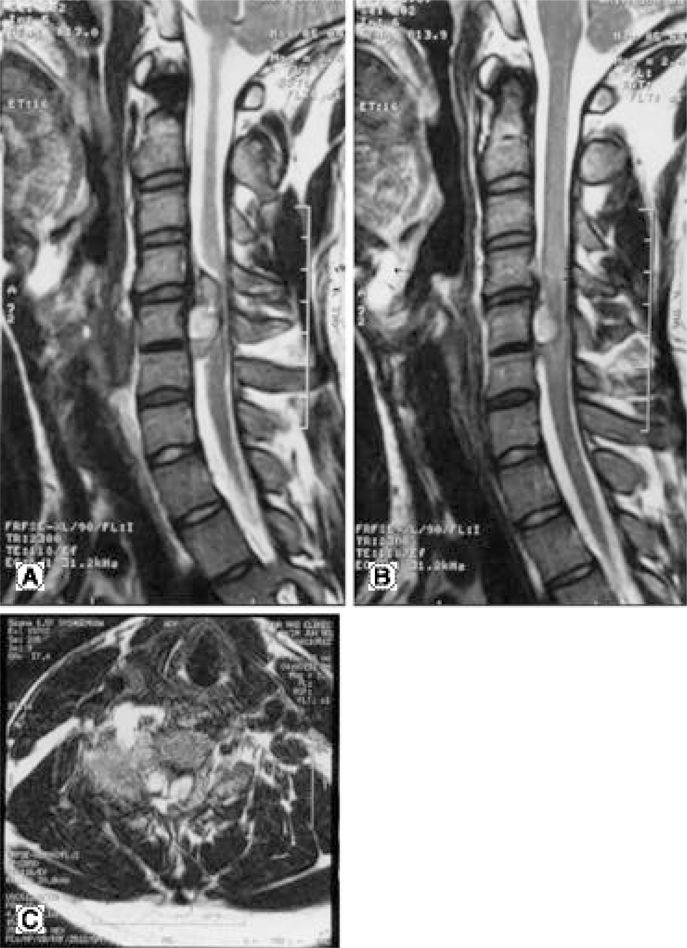
Fig. 2. (A, B) In the cervical MRI, it shows amorphous antevertebral ossification on 5-6 cervical spine and marked widening of anterior paravertebral soft tissue space, spinal cord compression by protruding mass.
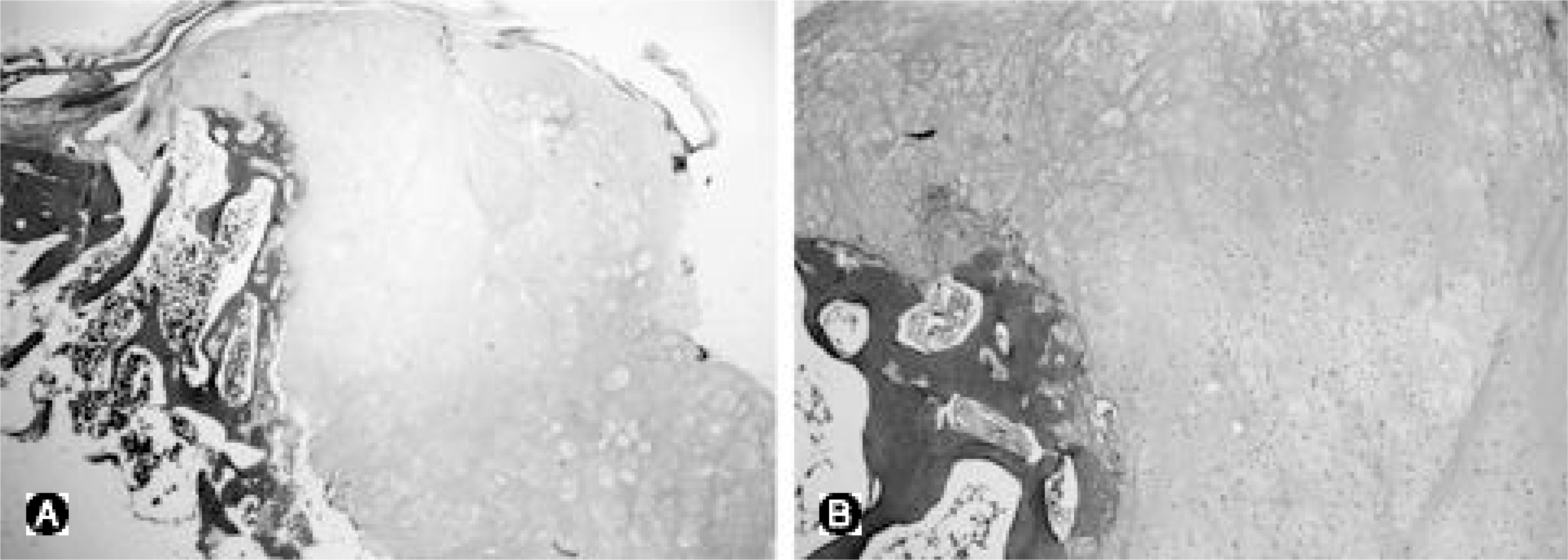
Fig. 3. (A,B) Histologically, the lesion contains very cellular cartilage, a proliferation of bizarre fibroblasts, and disorganized bone with spindle shaped fibroblasts in the trabecular spaces.
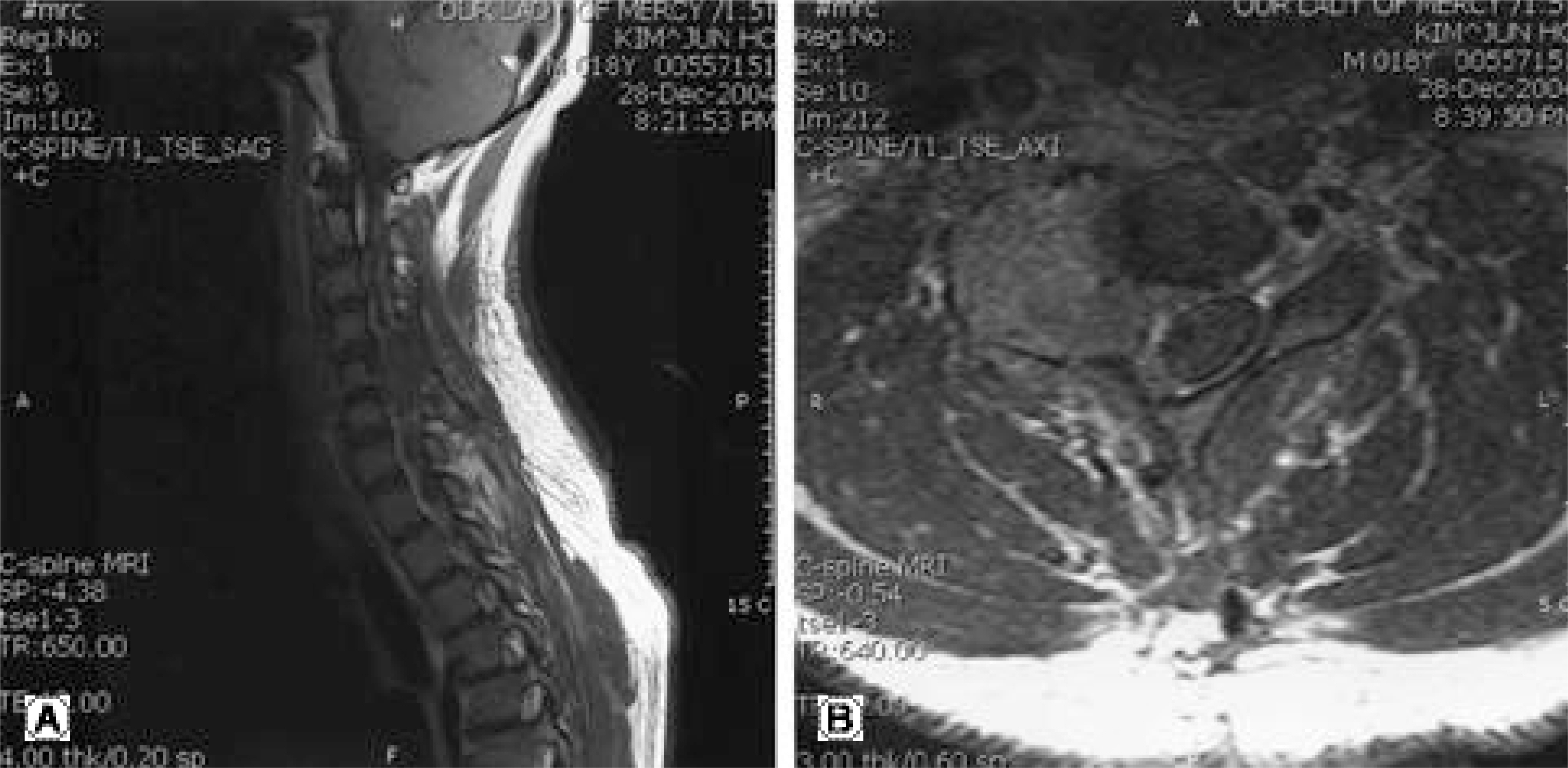
Fig. 4. (A,B) In the cervical MRI, it shows post-excision state of osteochondroma through cervical 4-6 level with small remaining mass. and also shows remarkable shift to left and compression of cord by postoperative hematoma.
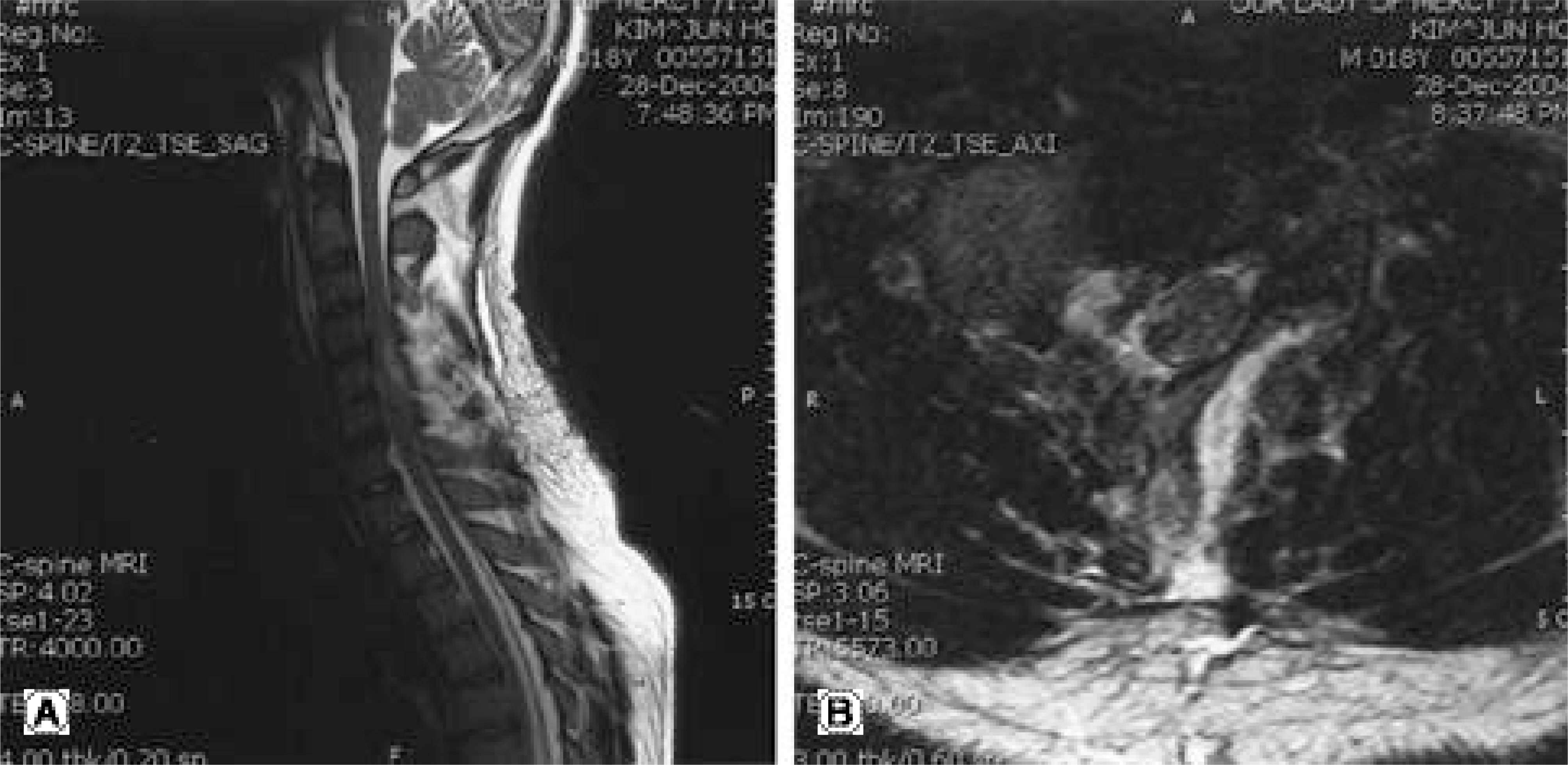
Fig. 5. (A,B) Contrast enhance cervical MRI images of above
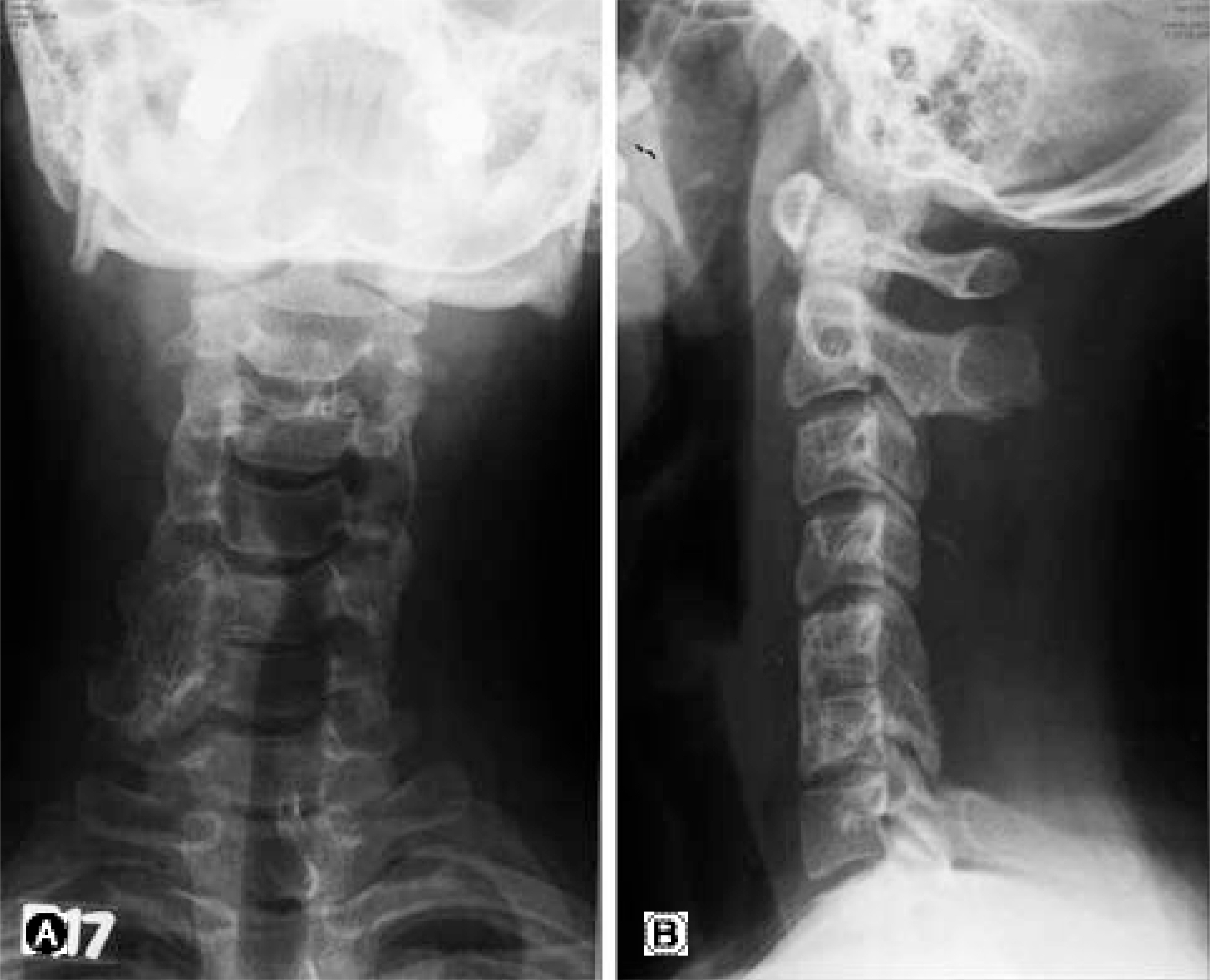
Fig. 6. In the post-operation plain X-ray, it shows removal of entire bony mass and decompressive total laminectomy of 3-6 cevical spine.
Reference
-
1). Jan Malat, Chat Virapongse, Alan Levine. solitary osteochondroma of the Spine. Case Report. Spine. 11(6):625–628. 1986.2). Prasad A, Renjen PN, Prasad ML, et al. Solitary spinal osteochondroma causing neural syndromes. Paraplegia. 30(9):678–680. 1992.
Article3). Raskas DS, Grazziano GP, Herzenberg JE, et al. Osteoid osteoma and osteoblastoma of the cervical spine. J Spinal Disord. 5(2):204–11. 1992.4). Cookes RS, Cumming WJ, Cowie RA. Osteochondroma of the cervical spine, Case report and review of the literature. Br J Neurosurg. 8(3):359–363. 1994.5). Morikawa M, Numaguchi Y, Soliman JA. Osteochondroma of the cervical spine. MR findings. Clin Imaging. 19(4):275–278. 1995.
Article6). Mermer , Matthew J. Gupta, Munish C.; Salamon, Peter B.; Benson, Daniel R. Thoracic Vertebral Body Exostosis as a Cause of Myelopathy in a Patient With Hereditary Multiple Exostoses. Journal of Spinal Disorders & Tech niques. 15(2):144–148. 2002.7). Arasil, Ertekin, Erdem, Atilla, Yuceer, Nurullah. Osteochondroma of the Upper Cervical Spine. A Case Report. Spine. 21(4):516–8. 1996.8). George B, Atallah A, Laurin C, et al. Cervical osteochondroma(C2 level) with vertebral artery occlusion and second cervical nerve root irritation. Surg Neurol, 9:. 31(6):459–464. 1989.9). Sheikhrezaii AR, Shariat S, Saberi H. Isolated Cervical Osteochondroma Manifesting as Spasmodic Torticollis. a Case Rreport. Irn J Med Sci. 25(1&2):87–89. 2000.10). Huvos AG. Bone tumors, Diagnosis Treatment and Prognosis. 2nd ed.Philadelphia: W.B. Ssunders Co.;1991.11). Jackson A, Hughes D, St Clair Forbes W, et al. A case of osteochondroma of the cervical spine. Skeletal Radiol. 24(3):235–237. 1995.
Article12). Eaton BA, Kettner NW, Essman JB. Solitary osteochondroma of the cervical spine. J Manipulative Physiol Ther. 18(4):250–253. 1995.13). Khosla A, Martin DS, Awwad EE. The solitary intraspinal vertebral Osteochondroma. Spine. 24(1):77–81. 1999.
Article
- Full Text Links
-
- Actions
-
Cited
- CITED
-
- Close
- Share
-
- Similar articles
-
- Dysphagia Caused by Osteochondroma of the Cervical Vertebral Body: A Case Report
- A Case of Osteochondroma which Arised form Right Side lamina of 5th Lumbar Vertebra
- Cervical Cord Injury Associated with Osteochondroma Involving Cervical Lamina: Case Report
- Solitary Osteochondroma of the Atlas with Cervical Cord Compresion: Case Report
- Osteochondroma of the Cervical Spine: A Case Report
