Revision of Atlantoaxial Fusion using Segmental Screw Fixation: Experience in Bilateral Posterior Arch Fracture of the Atlas Complicating Atlantoaxial Halifax Clamp Fixation: A Case Report
- Affiliations
-
- 1Department of Orthopaedic Surgery, Seoul Medical Center, Seoul, Korea.
- 2Department of Orthopaedic Surgery, Seoul National University College of Medicine, Seoul, Korea. ortho@hananet.net
- KMID: 1941651
- DOI: http://doi.org/10.4184/jkss.2007.14.3.187
Abstract
- A fracture of the posterior arch of the atlas is a rare complication of Halifax clamp fixation for atlantoaxial fusion. To the best of our knowledge, there is only one case reported reporting the English literature. Revision for this condition is challenging because of the difficulty in the surgical approach, internal fixation, and fusion. We report a case of bilateral fractures and nonunion of the posterior arch of the atlas and atlantoaxial nonunion after an atlantoaxial fusion procedure using Halifax clamp fixation, which resulted in persistent atlantoaxial instability and progressive myelopathy. Segmental screw fixation was performed using C1 lateral mass screws and C2 subarticular screws, along with intraarticular and extraarticular atlantoaxial inter-facet fusion.
MeSH Terms
Figure
-

Fig. 1. Preoperative lateral radiographs in flexion (A) and extension (B) show definite atlantoaxial instability and os odontoideum. In flexion (A), increased atlanto-dens interval (black arrow), nonunion between the bone graft and the axis (black arrowhead), and fracture of the posterior arch of the atlas (white arrowhead) are seen clear-ly.

Fig. 2. Preoperative CT scan shows bilateral fractures of the posterior arch of the atlas at just lateral to the Halifax clamps (black arrowheads).
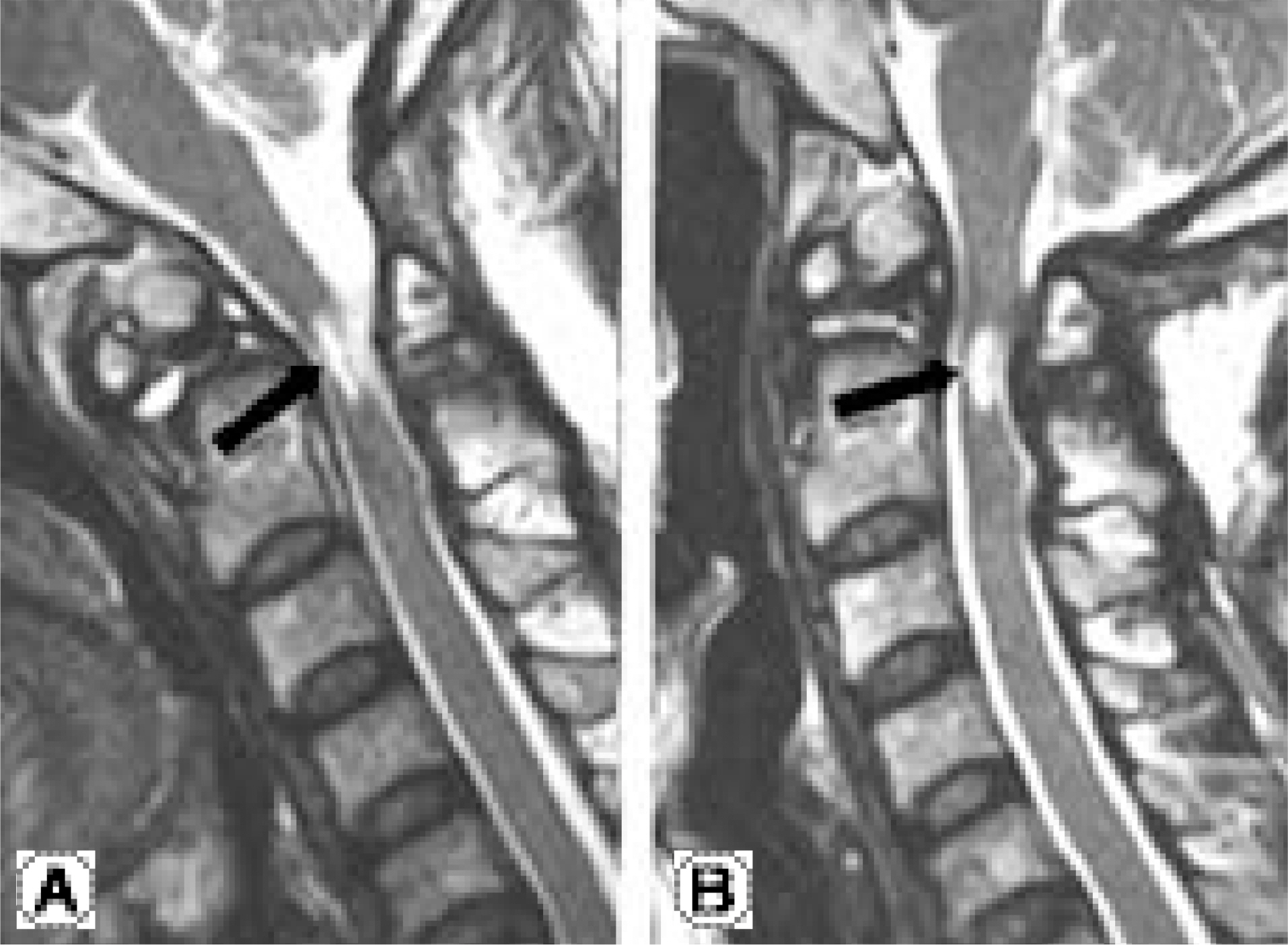
Fig. 3. Preoperative T2-weighted sagittal MRI in flexion (A) and extension (B) show atrophy of the spinal cord and high signal intensity area (black arrows) which is thought to be a syrinx.

Fig. 4. A photograph taken after curettage of the right atlantoaxial facet joint is shown. Subchondral bones of the facet joint (black arrowheads) are exposed. The white area between the two subchondral bones is the intraarticular cartilages reaming in the front.

Fig. 5. Radiographs taken 6 months after the operation are shown. (A) An open-mouth anteroposterior view show bone graft in the atlantoaxial facet joint (black arrowheads). (B) A lateral view shows good union of the extraarticular facet fusion (black arrow). Note that there is no bone in the corresponding area in preoperative lateral views (Fig. 1)
Reference
-
1). Aldrich EF, Weber PB, Crow WN. Halifax interlaminar clamp for posterior cervical fusion: A longterm followup review. J Neurosurg. 1993; 78:702–708.
Article2). Yeom JS, Won JH, Park SK, et al. The subarticular screw: A new trajectory for the C2 screw. J Korean Soc Spine Surg. 2006; 13:75–80.
Article3). Brooks AL, Jenkins EB. Atlantoaxial arthrodesis by the wedge compression method. J Bone Joint Surg. 1978; 60A:279–284.
Article4). Maniker AH, Schulder M, Duran HL. Halifax clamps: Efficacy and complications in posterior cervical stabilization. Surg Neurol. 1995; 43:140–146.
Article5). Moskovich R, Crockard HA. Atlantoaxial arthrodesis using interlaminar clamps. An improved technique. Spine. 1992; 17:261–267.6). Statham P, O'Sullivan M, Russell T. The Halifax interlaminar clamp for posterior cervical fusion: Initial experince in the United Kingdom. Neurosurgery. 1993; 32:396–399.7). Foley KT. Atlantoaxial fixation. (in. Papadopoulos SM, editor. Manual of cervical spine internal fixation. 1st ed.Philadelphia, Lippincott: Williams & Wilkins;p. 101. 2004.8). Gallie WE. Fracture and dislocation of the cervical spine. Am J Surg. 1939; 46:495–499.9). Goel A, Desai KI, Muzumdar DP. Atlantoaxial fixation using plate and screw method: a report of 160 treated patients. Neurosurgery. 2002; 51:1351–1357.
Article10). Suk KS, Kim KT, Lee SH. C1-2 C1-2 Transarticular Screw Fixation as a Revision Surgery for Failed C1-2 Fusion. J Korean Soc Spine Surg. 2002; 9:251–256.11). Lee KY, Lee MJ, Kim W. Double plate occipitocervical fusion after failed posterior fusion C1-2 with wiring. J Korean Soc Spine Surg. 2004; 11:121–124.
- Full Text Links
-
- Actions
-
Cited
- CITED
-
- Close
- Share
-
- Similar articles
-
- Old Atlantoaxial Rotary Subluxation Associated with High-riding Vertebral Arteries: Arthrodesis Using C1 Lateral Mass Screws and C2 Laminar Screws: A Case Report
- Application of C1-C3 Halifax Interlaminar Clamps in Addition to C1-C2 Cable Fixation
- C1-2 Transarticular Screw Fixation as a Revision Surgery for Failed C1-2 Fusion: Case Report
- C1-2 Fixation Using Polyaxial Screws and Rods Assisted by Computer Simulation for Revision of Failed Posterior Fusion: A Technical Report
- Clinical and Biomechanical Analysis of Transarticular Screw Fixation for Atlantoaxial Instability
