Uterine Adenomyosis Which Developed from Hypoplastic Uterus in Postmenopausal Woman with Mayer-Rokitansky-Kuster-Hauser Syndrome: A Case Report
- Affiliations
-
- 1Department of Obstetrics and Gynecology, Inje University Haeundae Paik Hospital, Busan, Korea. jyimd@lycos.co.kr
- 2Department of Pathology, Inje University Haeundae Paik Hospital, Busan, Korea.
- KMID: 1939537
- DOI: http://doi.org/10.6118/jmm.2013.19.3.135
Abstract
- Mayer-Rokitansky-Kuster-Hauser syndrome (MRKHS) is characterized by vaginal agenesis with variable Mullerian duct abnormalities. We report here a case of uterine adenomyosis which developed from a hypoplastic uterus in a patient with MRKHS. A 55-year-old postmenopausal woman visited a university hospital for pelvic mass. She had underwent vaginoplasty via the McIndoe procedure for MRKHS at 15 years of age. Pelvic magnetic resonance imaging showed a 5.4 x 4.8 x 4.7 cm mass suspicious for a uterine myoma. She received total abdominal hysterectomy with bilateral salpingo-oophorectomy, and neither the cervix nor endometrium was found grossly in the surgical specimen. The final histologic diagnosis was uterine adenomyosis.
MeSH Terms
Figure
-

Fig. 1 Pelvis magnetic resonance imaging. (A) Uterine mass, coronal view. (B) Uterine mass, sagittal view.

Fig. 2 Laparotomic view of the uterine mass.
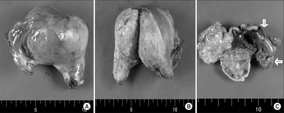
Fig. 3 Gross findings of the specimen. Multinodular uterus shows adenomyoma or myoma like features and the atrophic endometrum as well as the aplastic uterine cervix and vagina. White arrow points to the atrophic ovary and salpinx.
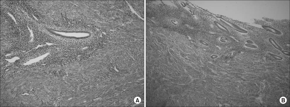
Fig. 4 Histologic findings from the resected specimen. (A) Uterine myometrium (H&E, × 200). Microscopically, the uterus shows typical adenomyosis features, such as multifocal atrophic endometrial glands and stroma in the background of proliferating smooth muscle cell bundles. (B) Uterine endometrium (H&E, × 100). The atrophoic endometrium (< 1 mm thickness) is seen in the lower segment of the uterus.
Reference
-
1. Fritz MA, Speroff L, editors. Clinical gynecologic endocrinology and infertility. 8th ed. Philadelphia, PA: Lippincott Williams & Wilkins;2010. p. 455–457. p. 612–613.2. Aittomäki K, Eroila H, Kajanoja P. A population-based study of the incidence of Mullerian aplasia in Finland. Fertil Steril. 2001; 76:624–625.3. Fedele L, Bianchi S, Frontino G, Ciappina N, Fontana E, Borruto F. Laparoscopic findings and pelvic anatomy in Mayer-Rokitansky-Kuster-Hauser syndrome. Obstet Gynecol. 2007; 109:1111–1115.4. Kim TH, Lee HH, Jeon DS, Park J. Laparoscopic resection of the rudimentary horn of a unicornuate uterus diagnosed by three-dimensional computed tomography. Med Case Stud. 2013; 4:9–12.5. McIndoe AH, Banister JB. An operation for the cure of congenital absence of the vagina. BJOG. 1938; 45:490–494.6. Rapkin AJ, Howe CN. Pelvic pain and dysmenorrhea. In : Berek JS, Novak E, editors. Berek & Novak's gynecology. 14th ed. Philadelphia, PA: Lippincott Williams & Wilkins;2007. p. 505–540.7. Enatsu A, Harada T, Yoshida S, Iwabe T, Terakawa N. Adenomyosis in a patient with the Rokitansky-Kuster-Hauser syndrome. Fertil Steril. 2000; 73:862–863.8. Yan CM, Mok KM. Uterine fibroids and adenomyosis in a woman with Rokitansky-Kuster-Hauser syndrome. J Obstet Gynaecol. 2002; 22:561–562.9. Nisolle M, Donnez J. Peritoneal endometriosis, ovarian endometriosis, and adenomyotic nodules of the rectovaginal septum are three different entities. Fertil Steril. 1997; 68:585–596.10. Chun S, Jeon GH, Cho HJ, Kim YM, Ji YI. Endometrial hyperplasia in myometrium of woman with uterine adenomyosis: a case report. J Reprod Endocrinol. 2012; 4:56–60.
- Full Text Links
-
- Actions
-
Cited
- CITED
-
- Close
- Share
-
- Similar articles
-
- A Case of Uterine Leiomyomas in both Rudimentary Uterine Horns in a Woman with the Mayer-Rokitansky-Kuster-Hauser Syndrome
- MR Findings of Mayer-Rokitansky-Kuster-Hauser Syndrome: Two Cases Report
- A Case of Mayer-Rokitansky-Kuster-Hauser Syndrome Combined with Single Pelvic Kidney
- A case of ovarian cancer developed in Mayer-Rokitansky-Kuster-Hauser syndrome: Innate carcinogenesis of ovarian cancer
- A Case of Mayer-Rokitansky-Kuster-Hauser (MRKH) Syndrome with Amenorrhea and Sexual Precosity
