Acute cholecystitis associated with Clonorchis sinensis infection
- Affiliations
-
- 1Department of Surgery, Digestive Disease Research Institute and Institute of Medical Science, Wonkwang University College of Medicine, Iksan, Korea. east1st@wku.ac.kr
- 2Department of Pathology, Digestive Disease Research Institute and Institute of Medical Science, Wonkwang University College of Medicine, Iksan, Korea.
- KMID: 1882839
- DOI: http://doi.org/10.4174/astr.2014.87.2.104
Abstract
- Clonorchis sinensis is one of the most common causes of trematodiasis that is caused by the ingestion of raw fish contaminated with infective cysts. The adult flukes are predominantly present in the intrahepatic bile ducts, but occasionally they may be found in the pancreatic duct and extrahepatic bile ducts. The clinical manifestations depend on the number of flukes, the period of infestation, and complications such as pericholangitic abscess, cholangitis, bile duct stones, and cholangiocarcinoma. However, primary acute cholecystitis associated with C. sinensis infection is extremely rare. Herein, we report on a case of primary acute cholecystitis associated with C. sinensis infection.
Keyword
MeSH Terms
Figure
-
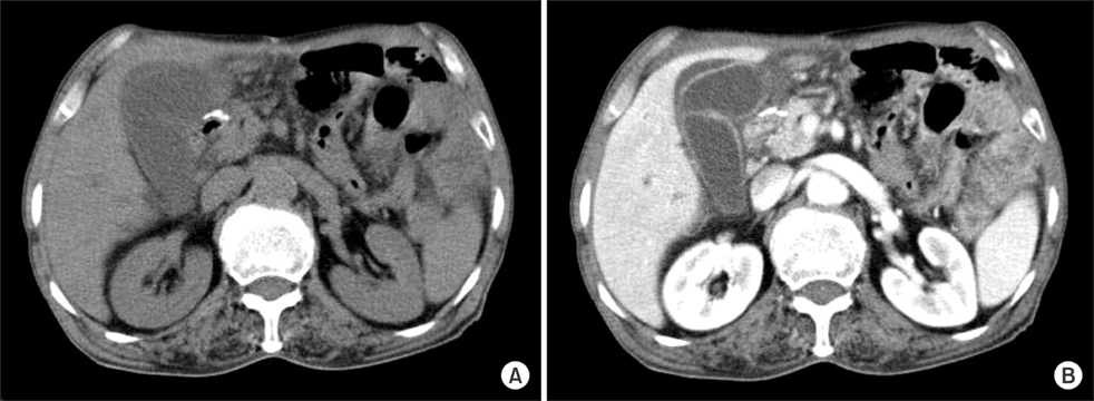
Fig. 1 Axial unenhanced (A) and contrast-enhanced abdominal computed tomography images (B) show a distended gallbladder with wall thickening and pericholecystic fluid collection. It also shows minimal dilatation of the peripheral intrahepatic bile ducts.
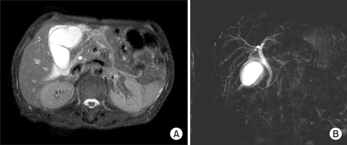
Fig. 2 MR cholangiography shows a distended gallbladder, pericholecystic fluid collection, and mild extrahepatic bile duct dilation without obstructive lesions.
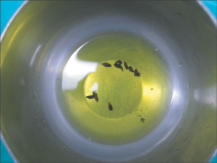
Fig. 3 Operative finding shows several flukes removed from the cystic duct and gallbladder.
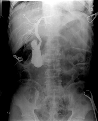
Fig. 4 Operative cholangiography shows neither bile duct dilatation nor obstructive lesions.
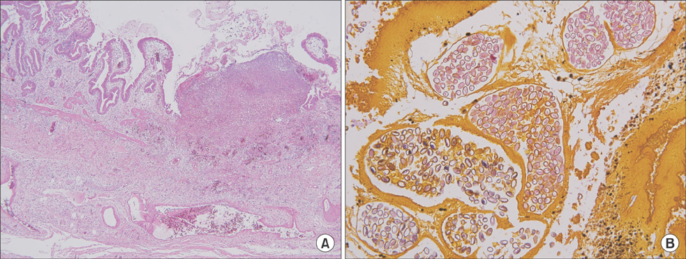
Fig. 5 Histopathological examinations show a severe inflammatory mucosal change of the gallbladder (A: H&E, ×40) and ovum (B: H&E, ×200), which is confirmed by the presence of Clonorchis sinensis.
Reference
-
1. Hong ST, Fang Y. Clonorchis sinensis and clonorchiasis, an update. Parasitol Int. 2012; 61:17–24.2. Qiao T, Ma RH, Luo XB, Luo ZL, Zheng PM. Cholecystolithiasis is associated with Clonorchis sinensis infection. PLoS One. 2012; 7:e42471.3. Stunell H, Buckley O, Geoghegan T, Torreggiani WC. Recurrent pyogenic cholangitis due to chronic infestation with Clonorchis sinensis (2006: 8b). Eur Radiol. 2006; 16:2612–2614.4. Shin HR, Oh JK, Masuyer E, Curado MP, Bouvard V, Fang YY, et al. Epidemiology of cholangiocarcinoma: an update focusing on risk factors. Cancer Sci. 2010; 101:579–585.5. Lai CH, Chin C, Chung HC, Liu H, Hwang JC, Lin HH. Clonorchiasis-associated perforated eosinophilic cholecystitis. Am J Trop Med Hyg. 2007; 76:396–398.6. Kim EM, Kim JL, Choi SY, Kim JW, Kim S, Choi MH, et al. Infection status of freshwater fish with metacercariae of Clonorchis sinensis in Korea. Korean J Parasitol. 2008; 46:247–251.7. Choi D, Hong ST. Imaging diagnosis of clonorchiasis. Korean J Parasitol. 2007; 45:77–85.8. Kim GH, Kang DH, Kim TO, Song GA, Heo J, Cho M, et al. MRCP and ERCP findings in clonorchiasis. Gastrointest Endosc. 2006; 64:439–440.9. Choi D, Lim JH, Lee KT, Lee JK, Choi SH, Heo JS, et al. Gallstones and Clonorchis sinensis infection: a hospital-based case-control study in Korea. J Gastroenterol Hepatol. 2008; 23(8 Pt 2):e399–e404.
- Full Text Links
-
- Actions
-
Cited
- CITED
-
- Close
- Share
-
- Similar articles
-
- A Case of Clonorchis Sinensis Infection in the Gastric Pylorus Associated with Gastric Adenocarcinoma
- Clonorchis sinensis Infection Presenting as Acute Cholangitis and Acute Cholecystitis
- A case of eosinophilic cholecystitis with hepatitis associated with clonorchiasis
- The intensity of clonorchis sinensis infection by E.P.G. counts and the degree of abnormalities in laboratory tests
- Clonorchis sinensis in Kyungpook Province, Korea 1. Distribution and demonstration of the cercaria of Clonorchis sinensis from snail, Parafossarulus manchouricus Bourgigant
