Korean Circ J.
2010 Nov;40(11):581-586. 10.4070/kcj.2010.40.11.581.
Comparison of Plaque Composition in Diabetic and Non-Diabetic Patients With Coronary Artery Disease Using Multislice CT Angiography
- Affiliations
-
- 1Department of Internal Medicine, Busan St. Mary's Hospital, Busan, Korea.
- 2Department of Internal Medicine, Inje University College of Medicine, Haeundae Paik Hospital, Busan, Korea.
- 3Department of Internal Medicine, Inje University College of Medicine, Busan Paik Hospital, Busan, Korea. jsjang@medimail.co.kr
- KMID: 1826177
- DOI: http://doi.org/10.4070/kcj.2010.40.11.581
Abstract
- BACKGROUND AND OBJECTIVES
Plaque composition rather than degree of luminal narrowing may be predictive of future coronary events in high risk patients. The purpose of this study was to compare degree of plaque burden and composition with multislice computed tomography (MSCT) angiography between diabetic and non-diabetic patients.
SUBJECTS AND METHODS
A total of 452 consecutive MSCT angiography examinations were performed between July 2007 and June 2009. Of these, the patients who underwent invasive coronary angiography were evaluated for the presence and type of atherosclerotic plaque and severity of luminal narrowing.
RESULTS
Ninety two (46 in the diabetic group and 46 in the non-diabetic group) patients underwent both MSCT angiography and invasive coronary angiography. Among them, 30 patients (65.2%) in the diabetic group and 26 patients (56.5%) in the non-diabetic group had significant coronary narrowing on MSCT angiography. Sixteen patients (34.8%) in the diabetic group and 15 patients (32.6%) in non-diabetic group underwent coronary angioplasty and stenting. Forty-two patients (93.3%) in the diabetic group and 39 patients (88.6%) in the non-diabetic group had multiple types of coronary plaque (p=0.485). MSCT angiography was similar to conventional coronary angiography in its ability to predict significant coronary artery disease in that the area under the curve was 0.88 (95% confidence interval, 0.81 to 0.95). Diabetic patients had more mixed plaque compared with non-diabetic patients.
CONCLUSION
Differences in coronary plaque composition between diabetic and non-diabetic patients can be determined noninvasively by MSCT angiography. In patients with diabetes, mixed plaque types contribute to the total plaque burden to a higher degree than in non-diabetic patients.
Keyword
MeSH Terms
Figure
-
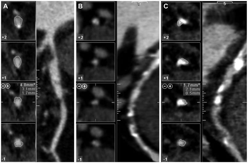
Fig. 1 Example of different plaque types as visually assigned on the multislice computed tomography angiography. A: non-calcified plaque. B: mixed plaque. C: calcified plaque.
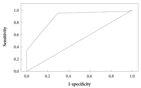
Fig. 2 Diagnostic performance of MSCT angiography. Receiver-operating-characteristic curve describing the diagnostic performance of MSCT angiography to identify coronary stenosis of 60% or more in culprit vessel. The area under the curve was 0.88 (95% confidence interval, 0.81 to 0.95). MSCT: multislice computed tomography.
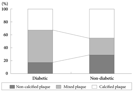
Fig. 3 Bar graph demonstrating the relative distribution of the different lesion types in diabetic and non-diabetic patients (p=0.035).
Reference
-
1. Achenbach S, Ulzheimer S, Baum U, et al. Noninvasive coronary angiography by retrospectively ECG-gated multi-slice spiral CT. Circulation. 2000. 102:2823–2828.2. Hoffmann U, Moselewski F, Cury RC, et al. Predictive value of 16-slice multi-detector spiral computed tomography to detect significant obstructive coronary artery disease in patients at high risk for coronary artery disease: patient-versus segment-based analysis. Circulation. 2004. 110:2638–2643.3. Oh HJ, Kwon K, Park SH, et al. CT coronary angiography using multidetector computed tomography in coronary artery disease : a comparative study to quantitative coronary angiography. Korean Circ J. 2004. 34:1167–1173.4. Achenbach S, Moselewski F, Ropers D, et al. Detection of calcified and non-calcified coronary atherosclerotic plaque by contrast-enhanced, submillimeter multi-detector spiral computed tomography: a segment based comparison with intravascular ultrasound. Circulation. 2004. 109:14–17.5. Kim SY, Kim KS, Lee YS, et al. Assessment of non-calcified coronary plaques using 64-slice computed tomography: comparison with intravascular ultrasound. Korean Circ J. 2009. 39:95–99.6. Cury RC, Pomerantsev EV, Ferencik M, et al. Comparison of the degree of coronary stenoses by multi-detector computed tomography versus by quantitative coronary angiography. Am J Cardiol. 2005. 96:784–787.7. Naghavi M, Libby P, Falk E, et al. From vulnerable plaque to vulnerable patient: a call for new definitions and risk assessment strategies: part I. Circulation. 2003. 108:1664–1672.8. Hoffmann U, Moselewski F, Nieman K, et al. Noninvasive assessment of plaque morphology and composition in culprit and stable lesions in acute coronary syndrome and stable lesions in stable angina by multi-detector computed tomography. J Am Coll Cardiol. 2006. 47:1655–1662.9. Schroeder S, Kopp AF, Baumbach A, et al. Noninvasive detection and evaluation of atherosclerotic coronary plaques with multi-slice computed tomography. J Am Coll Cardiol. 2001. 37:1430–1435.10. Ibebuogu UN, Nasir K, Gopal A, et al. Comparison of atherosclerotic plaque burden and composition between diabetic and non diabetic patients by non invasive CT angiography. Int J Cardiovasc Imaging. 2009. 25:717–723.11. Nicholls SJ, Tuzcu EM, Crowe T, et al. Relationship between cardiovascular risk factors and atherosclerotic disease burden measured by intravascular ultrasound. J Am Coll Cardiol. 2006. 47:1967–1975.12. Wayhs R, Zelinger A, Raggi P. High coronary artery calcium scores pose an extremely elevated risk for hard events. J Am Coll Cardiol. 2002. 39:225–230.13. Kim D, Choi SY, Choi EK, et al. Distribution of coronary artery calcification in an asymptomatic Korean population: association with risk factors of cardiovascular disease and metabolic syndrome. Korean Circ J. 2008. 38:29–35.14. Herlitz J, Karlson BW, Lindqvist J, Sjolin M. Rate and mode of death during five years of follow-up among patients with acute chest pain with and without a history of diabetes mellitus. Diabet Med. 1998. 15:308–314.15. Mann JM, Davies MJ. Vulnerable plaque: relation of characteristics to degree of stenosis in human coronary arteries. Circulation. 1996. 94:928–931.16. Giroud D, Li JM, Urban P, Meier B, Rutishauer W. Relation of the site of acute myocardial infarction to the most severe coronary arterial stenosis at prior angiography. Am J Cardiol. 1992. 69:729–732.17. Davies MJ, Thomas AC. Plaque fissuring: the cause of acute myocardial infarction, sudden ischaemic death, and crescendo angina. Br Heart J. 1985. 53:363–373.18. Kopp AF. Angio-CT: heart and coronary arteries. Eur J Radiol. 2003. 45:Suppl 1. S32–S36.19. Escolar E, Weigold G, Fuisz A, Weissman NJ. New imaging techniques for diagnosing coronary artery disease. CMAJ. 2006. 174:487–495.20. Carrascosa PM, Capunay CM, Garcia-Merletti P, Carrascosa J, Garcia MF. Characterization of coronary atherosclerotic plaques by multi-detector computed tomography. Am J Cardiol. 2006. 97:598–602.21. Hausmann D, Erbel R, Alibelli-Chemarin MJ, et al. The safety of intracoronary ultrasound: a multicenter survey of 2207 examinations. Circulation. 1995. 91:623–630.22. Pundziute G, Schuijf JD, Jukema JW, et al. Head-to-head comparison of coronary plaque evaluation between multislice computed tomography and intravascular ultrasound radiofrequency data analysis. JACC Cardiovasc Interv. 2008. 1:176–182.23. Kolodgie FD, Virmani R, Burke AP, et al. Pathologic assessment of the vulnerable human coronary plaque. Heart. 2004. 90:1385–1391.24. Rodriguez-Granillo GA, Garcia-Garcia HM, McFadden EP, et al. In vivo intravascular ultrasound-derived thin-cap fibroatheroma detection using ultrasound radiofrequency data analysis. J Am Coll Cardiol. 2005. 46:2038–2042.25. Pundziute G, Schuijf JD, Jukema JW, et al. Prognostic value of multislice computed tomography coronary angiography in patients with known or suspected coronary artery disease. J Am Coll Cardiol. 2007. 49:62–70.26. Pundziute G, Schuijf JD, Jukema JW, et al. Noninvasive assessment of plaque characteristics with multi-slice computed tomography coronary angiography in symptomatic diabetic patients. Diabetes Care. 2007. 30:1113–1119.27. Hambly RI, Sherman L, Mehta J, et al. Reappraisal of the role of the diabetic state in coronary artery disease. Chest. 1976. 70:251–257.28. Springer I, Dewey M. Comparison of multislice computed tomography with intravascular ultrasound for detection and characterization of coronary artery plaques: a systematic review. Eur J Radiol. 2009. 71:275–282.
- Full Text Links
-
- Actions
-
Cited
- CITED
-
- Close
- Share
-
- Similar articles
-
- Diabetes Mellitus and Coronary Angiography
- Cardiac CT
- Coronary CT Angiography
- Assessment of Coronary Stenosis Using Coronary CT Angiography in Patients with High Calcium Scores: Current Limitations and Future Perspectives
- Comparison of Endothelium-Dependent Vasodilation According to the Presence of Diabetes in Coronary Artery Disease
