Brain Neurorehabil.
2014 Sep;7(2):93-100. 10.12786/bn.2014.7.2.93.
Functional Neuroanatomy of Brain Stem
- Affiliations
-
- 1Department of Rehabilitation Medicine, Seoul Asan Medical Center, Univeristy of Ulsan College Medicine, Korea. dykimsmart@gmail.com
- KMID: 1797641
- DOI: http://doi.org/10.12786/bn.2014.7.2.93
Abstract
- The brain stem consists of medulla oblongta, pons and midbrain. It is sited in posterior cranial fossa. It contains numerous intrinsic neuron cell bodies and their processes, some of which are the brain stem homologues of spinal neuronal groups. These include the sites of termination and cells of origin of axons that enter or leave the brain stem through the cranial nerves. Cranial nerves provide sensory, motor and autonomic innervations of structures that are mostly in the head and neck. The reticular formation is an extensive network of neurons that extends throughout the length of brain stem and is continuous rostrally to diencephalon and caudally to its spinal counterpart. Clinically, damage to the brain stem is often devastating and life threatening. This is because it is a structurally and functionally compact region. Therefore, it is important to build basic knowledge about neuroanatomy of brain stem.
Keyword
MeSH Terms
Figure
-

Fig. 1 Cranial nerve nuclei.
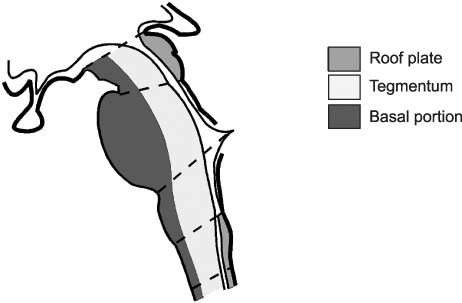
Fig. 2 Basic structure of brain stem.
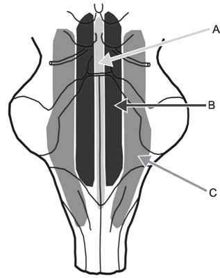
Fig. 3 Subdivision of reticular formation. (A) Raphe nuclei and paramedian group. (B) Medial group. (C) Lateral group.
Reference
-
1. Hendelman WJ. Atlas of Functional Neuroanatomy. Brainstem Histology. 2nd ed. Taylor & Francis;2006. p. 172–197.2. Blumenfeld H. Neuroanatomy through Clinical Cases. Brainstem I: Surface Anatomy and Cranial Nerves. 2nd ed. Sunderland, Massachusetts: Sinauer Associates, Inc.;2010.3. Afifi AK, Bergman RA. Functional Neuroanatomy. Medulla Oblonta: Large Medical Books/McGraw-Hill;2005. p. 78–97.4. Afifi AK, Bergman RA. Functional Neuroanatomy. Pons. Large Medical Books/McGraw-Hill;2005. p. 103–122.5. Afifi AK, Bergman RA. Functional Neuroanatomy. Midbrain. Large Medical Books/McGraw-Hill;2005. p. 129–149.6. Mancall EL, Brock DG. Gray's Clinical Neuroanatomy, The Anatomic Basis for Clinical Neuroscience. Brain Stem. Elsevier Saunders;2010. p. 157–184.
- Full Text Links
-
- Actions
-
Cited
- CITED
-
- Close
- Share
-
- Similar articles
-
- Suggestions on the Proper Improvement of the Neuroanatomy Lab - Based in the Survey Analysis on the Online Sectional Neuroanatomy Lab Lecture to the Neuroanatomy Class -
- Depression and the Frontal Lobe
- Functional Neuroanatomy for Posture and Gait Control
- Neuroanatomy of Unilateral Neglect Syndrome
- Information processing and its neuroanatomy in schizophrenia
