J Korean Med Sci.
2014 Jun;29(6):751-757. 10.3346/jkms.2014.29.6.751.
Overview of Surgical Scar Prevention and Management
- Affiliations
-
- 1Department of Plastic and Reconstructive Surgery, Keimyung University School of Medicine, Daegu, Korea.
- 2Well Plastic Surgery Clinic, Seoul, Korea. handson@dsmc.or.kr
- KMID: 1796934
- DOI: http://doi.org/10.3346/jkms.2014.29.6.751
Abstract
- Management of incisional scar is intimately connected to stages of wound healing. The management of an elective surgery patient begins with a thorough informed consent process in which the patient is made aware of personal and clinical circumstances that cannot be modified, such as age, ethnicity, and previous history of hypertrophic scars. In scar prevention, the single most important modifiable factor is wound tension during the proliferative and remodeling phases, and this is determined by the choice of incision design. Traditional incisions most often follow relaxed skin tension lines, but no such lines exist in high surface tension areas. If such incisions are unavoidable, the patient must be informed of this ahead of time. The management of a surgical incision does not end when the sutures are removed. Surgical scar care should be continued for one year. Patient participation is paramount in obtaining the optimal outcome. Postoperative visits should screen for signs of scar hypertrophy and has a dual purpose of continued patient education and reinforcement of proper care. Early intervention is a key to control hyperplastic response. Hypertrophic scars that do not improve by 6 months are keloids and should be managed aggressively with intralesional steroid injections and alternate modalities.
MeSH Terms
Figure
-

Fig. 1 Langer's lines.
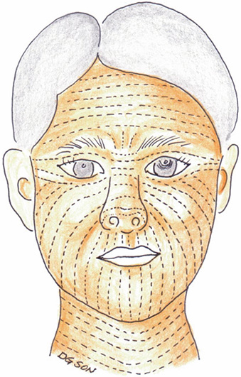
Fig. 2 Resting static tension lines on the face.
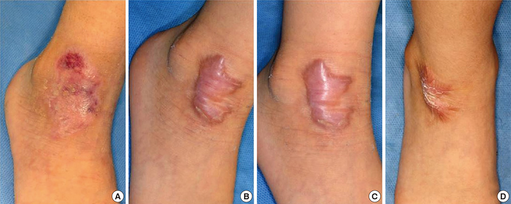
Fig. 3 Serial photographs of a healing wound from abrasion over the ankle. (A) At full epithelialization, (B) 3 months, (C) 6 months, and (D) 12 months from abrasion.
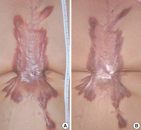
Fig. 4 Steroid injection in keloid. (A) Keloids over the sternum in a 62-yr-old female patient after open heart operation. (B) The keloid responded well after three injections of intralesional steroid.
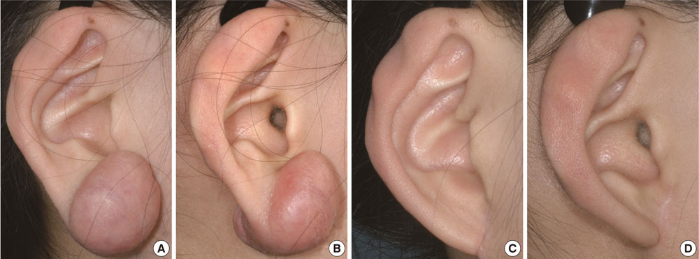
Fig. 5 Radiation therapy after keloid excision. (A, B) Photographic views of a 15-yr-old teenage girl with keloids from ear piercing. (C, D) One year after surgical excision and radiation therapy.
Reference
-
1. Almine JF, Wise SG, Weiss AS. Elastin signaling in wound repair. Birth Defects Res C Embryo Today. 2012; 96:248–257.2. Bae SH, Bae YC, Nam SB, Choi SJ. A skin fixation method for decreasing the influence of wound contraction on wound healing in a rat model. Arch Plast Surg. 2012; 39:457–462.3. Larson BJ, Longaker MT, Lorenz HP. Scarless fetal wound healing: a basic science review. Plast Reconstr Surg. 2010; 126:1172–1180.4. Kim HY, Kim JW, Park JH, Kim JH, Han YS. Personal factors that affect the satisfaction of female patients undergoing esthetic suture after typical thyroidectomy. Arch Plast Surg. 2013; 40:414–424.5. Toll EC, Loizou P, Davis CR, Porter GC, Pothier DD. Scars and satisfaction: do smaller scars improve patient-reported outcome? Eur Arch Otorhinolaryngol. 2012; 269:309–313.6. Chu HJ, Son DG, Kwon SY, Kim JH, Han KH. Characteristics of wound contraction according to the shape and antomical regions of the wound in porcine model. J Korean Soc Plast Reconstr Surg. 2011; 38:576–584.7. Wilhelmi BJ, Blackwell SJ, Phillips LG. Langer's lines: to use or not to use. Plast Reconstr Surg. 1999; 104:208–214.8. Borges AF. Relaxed skin tension lines (RSTL) versus other skin lines. Plast Reconstr Surg. 1984; 73:144–150.9. Borges AF. Relaxed skin tension lines. Dermatol Clin. 1989; 7:169–177.10. Bush JA, McGrouther DA, Young VL, Herndon DN, Longaker MT, Mustoe TA, Ferguson MW. Recommendations on clinical proof of efficacy for potential scar prevention and reduction therapies. Wound Repair Regen. 2011; 19:s32–s37.11. Kang BS, Na YC, Jin YW. Comparison of the wound healing effect of cellulose and gelatin: an in vivo study. Arch Plast Surg. 2012; 39:317–321.12. Shin HS, Oh HY. The effect of platelet-rich plasma on wounds of OLETF rats using expression of matrix metalloproteinase-2 and -9 mRNA. Arch Plast Surg. 2012; 39:106–112.13. Atkinson JA, McKenna KT, Barnett AG, McGrath DJ, Rudd M. A randomized, controlled trial to determine the efficacy of paper tape in preventing hypertrophic scar formation in surgical incisions that traverse Langer's skin tension lines. Plast Reconstr Surg. 2005; 116:1648–1656.14. Verhaegen PD, van Zuijlen PP, Pennings NM, van Marle J, Niessen FB, van der Horst CM, Middelkoop E. Differences in collagen architecture between keloid, hypertrophic scar, normotrophic scar, and normal skin: an objective histopathological analysis. Wound Repair Regen. 2009; 17:649–656.15. Bran GM, Goessler UR, Hormann K, Riedel F, Sadick H. Keloids: current concepts of pathogenesis (review). Int J Mol Med. 2009; 24:283–293.16. Juckett G, Hartman-Adams H. Management of keloids and hypertrophic scars. Am Fam Physician. 2009; 80:253–260.17. Chang CC, Kuo YF, Chiu HC, Lee JL, Wong TW, Jee SH. Hydration, not silicone, modulates the effects of keratinocytes on fibroblasts. J Surg Res. 1995; 59:705–711.18. Borgognoni L. Biological effects of silicone gel sheeting. Wound Repair Regen. 2002; 10:118–121.19. Sawada Y, Sone K. Treatment of scars and keloids with a cream containing silicone oil. Br J Plast Surg. 1990; 43:683–688.20. Tang YW. Intra- and postoperative steroid injections for keloids and hypertrophic scars. Br J Plast Surg. 1992; 45:371–373.21. Rosen DJ, Patel MK, Freeman K, Weiss PR. A primary protocol for the management of ear keloids: results of excision combined with intraoperative and postoperative steroid injections. Plast Reconstr Surg. 2007; 120:1395–1400.22. Niessen FB, Spauwen PH, Schalkwijk J, Kon M. On the nature of hypertrophic scars and keloids: a review. Plast Reconstr Surg. 1999; 104:1435–1458.23. Bouzari N, Davis SC, Nouri K. Laser treatment of keloids and hypertrophic scars. Int J Dermatol. 2007; 46:80–88.24. Parrett BM, Donelan MB. Pulsed dye laser in burn scars: current concepts and future directions. Burns. 2010; 36:443–449.25. Oluwasanmi JO. Keloids in the African. Clin Plast Surg. 1974; 1:179–195.26. Appleton I, Brown NJ, Willoughby DA. Apoptosis, necrosis, and proliferation: possible implications in the etiology of keloids. Am J Pathol. 1996; 149:1441–1447.27. Ladin DA, Hou Z, Patel D, McPhail M, Olson JC, Saed GM, Fivenson DP. P53 and apoptosis alterations in keloids and keloid fibroblasts. Wound Repair Regen. 1998; 6:28–37.28. DiPietro LA. Angiogenesis and scar formation in healing wounds. Curr Opin Rheumatol. 2013; 25:87–91.29. Huang C, Akaishi S, Ogawa R. Mechanosignaling pathways in cutaneous scarring. Arch Dermatol Res. 2012; 304:589–597.30. Sarrazy V, Billet F, Micallef L, Coulomb B, Desmoulière A. Mechanisms of pathological scarring: role of myofibroblasts and current developments. Wound Repair Regen. 2011; 19:s10–s15.31. Son DG, Lee HG, Han KH, Kim JH. Radiation therapy following total keloidectomy; a preliminary report. J Korean Soc Plast Reconstr Surg. 2005; 32:717–722.32. Nanda S, Reddy BS. Intralesional 5-fluorouracil as a treatment modality of keloids. Dermatol Surg. 2004; 30:54–56.33. Tziotzios C, Profyris C, Sterling J. Cutaneous scarring: pathophysiology, molecular mechanisms, and scar reduction therapeutics Part II. Strategies to reduce scar formation after dermatologic procedures. J Am Acad Dermatol. 2012; 66:13–24.34. Occleston NL, Fairlamb D, Hutchison J, O'Kane S, Ferguson MW. Avotermin for the improvement of scar appearance: a new pharmaceutical in a new therapeutic area. Expert Opin Investig Drugs. 2009; 18:1231–1239.35. Jackson WM, Nesti LJ, Tuan RS. Mesenchymal stem cell therapy for attenuation of scar formation during wound healing. Stem Cell Res Ther. 2012; 3:20.36. Sung HM, Suh IS, Lee HB, Tak KS, Moon KM, Jung MS. Case reports of adipose-derived stem cell therapy for nasal skin necrosis after filler injection. Arch Plast Surg. 2012; 39:51–54.37. Yang JD, Choi DS, Cho YK, Kim TK, Lee JW, Choi KY, Chung HY, Cho BC, Byun JS. Effect of amniotic fluid stem cells and amniotic fluid cells on the wound healing process in a white rat model. Arch Plast Surg. 2013; 40:496–504.
- Full Text Links
-
- Actions
-
Cited
- CITED
-
- Close
- Share
-
- Similar articles
-
- Facelift surgery : the importance of incision design and scar prevention
- Prevention of Postsurgical Scars: Comparsion of Efficacy and Convenience between Silicone Gel Sheet and Topical Silicone Gel
- Surgical Treatment of Keloid Scars on the Ear: The Usefulness of the Fillet Flap
- An experimental study on prevention of postlaminectomy scar formation
- The fetal wound healing: a review
