Serial Ultrasound and Computed Tomography Findings of Torsion of Lipomatous Appendage of the Falciform Ligament in a Child Treated by Conservative Management
- Affiliations
-
- 1Department of Radiology, Ulsan University Hospital, University of Ulsan College of Medicine, Ulsan, Korea. drcsh@naver.com
- KMID: 1793896
- DOI: http://doi.org/10.3348/jksr.2015.72.5.368
Abstract
- Torsion of the lipomatous appendage of the falciform ligament is extremely rare, and most patients have previously been treated surgically. We reported a case of torsion of the lipomatous appendage of the falciform ligament in a child, diagnosed by ultrasound (US) and computed tomography (CT) and treated conservatively. Real-time US and CT can aid an accurate diagnosis, and follow-up US can aid in making appropriate decisions for conservative treatment.
Figure
-
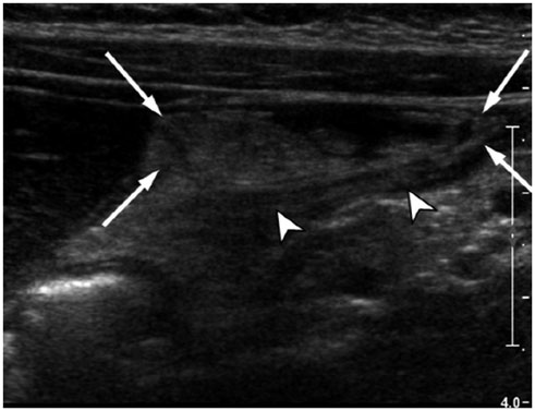
Fig. 1 Longitudinal US image of the right upper quadrant showed a non-compressible, heterogeneously hyperechoic, mass-like lesion (arrows) at the anterior of the falciform ligament (arrowheads) extending along with ligamentum teres. US = ultrasound
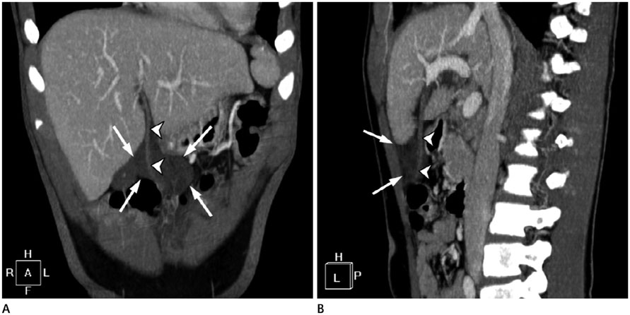
Fig. 2 Coronal (A) and sagittal (B) reformatted contrast-enhanced CT scans showed a heterogeneous, low attenuated, mass-like lesion (arrows) at the anterior of the falciform ligament (arrowheads). Note the lesion has a close relationship with the falciform ligament.

Fig. 3 In a follow-up US after 3 days, the characteristics of the lesion (arrows) was changed to homogeneous hyperechogenicity. US = ultrasound
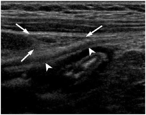
Fig. 4 In a follow-up US after 14 days, the previous lesion was almost completely resolved. The falciform ligament (arrowheads) appeared normal and scanty amount of fatty appendage (arrows) were noted. US = ultrasound
Reference
-
1. Brock JS, Pachter HL, Schreiber J, Hofstetter SR. Surgical diseases of the falciform ligament. Am J Gastroenterol. 1992; 87:757–758.2. Losanoff JE, Kjossev KT. Isolated gangrene of the round and falciform liver ligaments: a rare cause of peritonitis: case report and review of the world literature. Am Surg. 2002; 68:751–755.3. Crawford R, Anderson JR. Strangulated omental hernia of the falciform ligament. Br J Surg. 1985; 72:444.4. Webber CE Jr, Glanges E, Crenshaw CA. Falciform ligament. A possible twist? Arch Surg. 1977; 112:1264.5. Lloyd T. Primary torsion of the falciform ligament: computed tomography and ultrasound findings. Australas Radiol. 2006; 50:252–254.6. Coulier B, Cloots V, Ramboux A. US and CT diagnosis of a twisted lipomatous appendage of the falciform ligament. Eur Radiol. 2001; 11:213–215.7. Coulier B. Segmental omental infarction in childhood: a typical case diagnosed by CT allowing successful conservative treatment. Pediatr Radiol. 2006; 36:141–143.8. van Breda Vriesman AC, Lohle PN, Coerkamp EG, Puylaert JB. Infarction of omentum and epiploic appendage: diagnosis, epidemiology and natural history. Eur Radiol. 1999; 9:1886–1892.
- Full Text Links
-
- Actions
-
Cited
- CITED
-
- Close
- Share
-
- Similar articles
-
- Torsion of Fatty Appendage of Falciform Ligament Following Endoscopic Retrograde Cholangiopancreatography
- Abscess Formation Involving the Falciform Ligament and Ligamentum Teres
- Falciform Ligament Abscess after Omphalitis: Report of a Case
- Primary Abscess of the Falciform Ligament
- Minimally Invasive Treatment of Falciform Ligament Abscess in a 25-Day-Old Neonate: A Case Report
