Lymphangiomatosis Involving the Inferior Vena Cava, Heart, Pulmonary Artery and Pelvic Cavity
- Affiliations
-
- 1Department of Radiology, Soonchunhyang University Bucheon Hospital, Bucheon, Gyeonggi-do 420-767, Korea. dhk1107@hanmail.net
- 2Division of Cardiology, Department of Internal Medicine, Soonchunhyang University Bucheon Hospital, Bucheon, Gyeonggi-do 420-767, Korea.
- 3Department of Pathology, Soonchunhyang University Bucheon Hospital, Bucheon, Gyeonggi-do 420-767, Korea.
- 4Department of Thoracic Surgery, Soonchunhyang University Bucheon Hospital, Bucheon, Gyeonggi-do 420-767, Korea.
- 5Department of Anesthesiology and Pain Medicine, Asan Medical Center, Ulsan University College of Medicine, Seoul 138-736, Korea.
- KMID: 1787021
- DOI: http://doi.org/10.3348/kjr.2010.11.1.115
Abstract
- A 38-year-old woman who had undergone pelvic lymphangioma resection two months previously presented with cough and dyspnea. Transthoracic echocardiography and CT demonstrated the presence of a mixed cystic/solid component tumor involving the inferior vena cava, heart and pulmonary artery. Complete resection of the cardiac tumor was performed and lymphangioma was confirmed based on histopathologic examination. To the best of our knowledge, this is the first report of lymphangiomatosis with cardiac and pelvic involvement in the published clinical literature.
Keyword
MeSH Terms
Figure
-
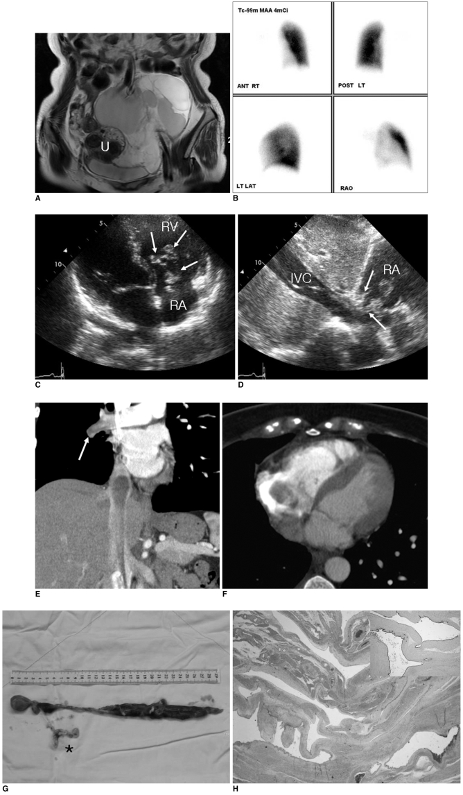
Fig. 1 Lymphangiomatosis in 38-year-old woman.A. Coronal T2-weighted image shows large lobulated cystic mass with many septations in pelvic cavity. Preserved uterus (U) without tumor involvement is seen.B. Perfusion lung scan with Tc-99m macroaggregated albumin shows perfusion defect in entire right lung, which was suspected to indicate complete occlusion of right pulmonary artery.C, D. Transthoracic echocardiographs show heterogeneous echogenic mass with cystic component and incomplete coaptation of tricuspid valve, which resulted in tricuspid regurgitation (arrows in C). Mass extended to inferior vena cava (IVC) (arrows in D) (RA = right atrium, RV = right ventricle).E, F. CT coronal (E) and axial (F) scans show non-enhanced mass with inferior vena cava and right atrial involvement, and hypoattenuating nodular lesion in right distal pulmonary artery (arrow in E).G. Gross specimen excised from inferior vena cava, right heart and right pulmonary artery was seen as an elongated, reddish, 29 cm long mass with web-like tumor extension in right pulmonary artery (asterisk).H. Hematoxylin & Eosin stained section of lesion shows dilated lymphatic channels with variable wall thicknesses. Based on immunohistochemical staining, tumor cells were positive for lymphatic vessel marker D2-40 (×40, insert).
Reference
-
1. Flörchinger B, Rümmele P, Lehane C, Schmid FX, Birnbaum DE. Mitral prolapse caused by lymphangioma. Thorac Cardiovasc Surg. 2005; 53:180–183. PMID: 15926101.
Article2. Kim SJ, Shin ES, Kim SW, Shin JK, Cheong JP, Kim YM, et al. A case of cardiac lymphangioma presenting as a cystic mass in the right atrium. Yonsei Med J. 2007; 48:1043–1047. PMID: 18159600.
Article3. Pennec PY, Blanc JJ. Cardiac lymphangioma: a benign cardiac tumour. Eur Heart J. 2006; 27:2913. PMID: 17145721.
Article4. Nataf P, Mestiri T, Martin de Lasalle E, Benomar M, Gandjbakhch I, Cabrol C. Pericardial hemolymphangioma. Apropos of a case. Arch Mal Coeur Vaiss. 1988; 81:1137–1140. PMID: 3143337.5. Kaji T, Takamatsu H, Noguchi H, Tahara H, Matsuda H, Nomura Y, et al. Cardiac lymphangioma: case report and review of the literature. J Pediatr Surg. 2002; 37:E32. PMID: 12378478.
Article6. Kemp JL, Kessler RM, Raizada V, Williamson MR. Case report. MR and CT appearance of cardiac hemangioma. J Comput Assist Tomogr. 1996; 20:482–483. PMID: 8626917.
Article7. Ozdemir H, Kocakoc E, Bozgeyik Z, Cobanoglu B. Recurrent retroperitoneal cystic lymphangioma. Yonsei Med J. 2005; 46:715–718. PMID: 16259073.
Article
- Full Text Links
-
- Actions
-
Cited
- CITED
-
- Close
- Share
-
- Similar articles
-
- Heart Related Disease: Chest CT Interpretation
- A case of intravascular leiomyomatosis extending to inferior vena cava, right heart, and pulmonary artery
- Transposition of inferior vena cava
- A case of hemiazygos continuation of a left inferior vena cava
- A Case of Congenital Absence of the Inferior Vena Cava
