A case of intravascular leiomyomatosis extending to inferior vena cava, right heart, and pulmonary artery
- Affiliations
-
- 1Department of Obstetrics and Gynecology, Asan Medical Center, University of Ulsan College of Medicine, Seoul, Korea. catgut1-0@hanmail.net
- 2Department of Thoracic and Cardiovascular Surgery, Asan Medical Center, University of Ulsan College of Medicine, Seoul, Korea.
- 3Department of Pathology, Asan Medical Center, University of Ulsan College of Medicine, Seoul, Korea.
- KMID: 1836768
- DOI: http://doi.org/10.5468/KJOG.2012.55.1.64
Abstract
- Intravascular leiomyomatosis is a rare entity of benign smooth muscle tumor invading into the lumen of veins. Although these tumors are histologically benign they sometimes extend into the cardiac cavity and can cause sudden death due to their incarceration into the artrioventricular orifice. We present a case of the intravascular leiomyomatosis originating from huge leiomyoma in the pelvic cavity and extending to the ovarian veins, the inferior vena cava, the right atrium, the right ventricle and the pulmonary artery. She underwent surgery by a single-stage o including laparotomy and venotomy without thoracotomy, and the tumor was successfully removed. Therefore, we report this case with a brief review of the literature.
MeSH Terms
Figure
-

Fig. 1 (A) Abdominal computed tomography (CT) showing mass (arrow) in the abdominal cavity. (B) Abdominal CT showing mass in the inferior vena cava. (C) Chest CT showing mass on right atrium and main pulmonary artery.

Fig. 2 (A) Huge mass extened to the right ovarian vein, the inferior vena cava, the right atrium and the pulmonary artery. The inferior vena cava is dilated as a 5 cm diameter. (B) Satinsky clamp grasp the inferior vena cava during repairing.
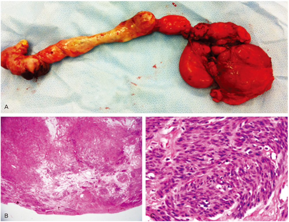
Fig. 3 (A) Intravenous leiomyomatosis. Intracaval worm-like, white mass, measuring 27 cm in length and abdominal mass, measuring 15 × 10 × 7 cm. (B) The resected tumor shows well develped and dilated blood vessels and is mixed cellular and acellular region (H&E, ×12.5). (C) A densely proliferation of small spindle cells with elongated nuclei (H&E, ×400).
Reference
-
1. Ariza A, Cerra C, Hahn IS, Shaw RK, Rigney B. Intravascular leiomyomatosis of the uterus. A case report. Conn Med. 1982. 46:700–703.2. Choi JY, Lee CJ, Kim WG, Cho SR, Shin HC. ntravenous leiomyomatosis with intracaval mass, intracardiac extension, and pulmonary metastasis: a case report. Korean J Obstet Gynecol. 2004. 47:1241–1245.3. Shida T, Yoshimura M, Chihara H, Nakamura K. Intravenous leiomyomatosis of the pelvis with reextension into the heart. Ann Thorac Surg. 1986. 42:104–106.4. Lo KW, Lau TK. Intracardiac leiomyomatosis. Case report and literature review. Arch Gynecol Obstet. 2001. 264:209–210.5. Lee KH, Bong JM, Shin MS, Kim JH, Shin EK, Jeon YB, et al. A case of intravenous leiomyomatosis extending into the right atrium. Korean Circ J. 2002. 32:825–828.6. Nogales FF, Navarro N, Martinez de Victoria JM, Contreras F, Redondo C, Herraiz MA, et al. Uterine intravascular leiomyomatosis: an update and report of seven cases. Int J Gynecol Pathol. 1987. 6:331–339.7. Kir G, Kir M, Gurbuz A, Karateke A, Aker F. Estrogen and progesterone expression of vessel walls with intravascular leiomyomatosis; discussion of histogenesis. Eur J Gynaecol Oncol. 2004. 25:362–366.8. Robboy SJ, Bentley RC, Butnor K, Anderson MC. Pathology and pathophysiology of uterine smooth-muscle tumors. Environ Health Perspect. 2000. 108:Suppl 5. 779–784.9. Kocaoglu M, Bulakbasi N, Ugurel MS, Ors F, Tayfun C, Ucoz T. Value of magnetic resonance imaging in the depiction of intravenous leiomyomatosis extending to the heart. J Comput Assist Tomogr. 2003. 27:630–633.10. Yang JH, Yoo JB, Park MI, Kim DS. Intravenous leiomyomatosis of the uterus: a case report. Korean J Obstet Gynecol. 1989. 32:997–1000.11. Arinami Y, Kodama S, Kase H, Tanaka K, Okazaki H, Maruyama Y. Successful one-stage complete removal of an entire intravenous leiomyomatosis in the heart, vena cava, and uterus. Gynecol Oncol. 1997. 64:547–550.12. Maurer G, Nanda NC. Two-dimensional echocardiographic identification of intracardiac leiomyomatosis. Am Heart J. 1982. 103:915–917.13. Esmaeilzadeh M, Tavokalli A, Yousefinia MA. Intracardiac leiomyomatosis. Iran Heart J. 2006. 7:61–66.
- Full Text Links
-
- Actions
-
Cited
- CITED
-
- Close
- Share
-
- Similar articles
-
- Three Cases of Intracardiac Leiomyomatosis with Very Long-term Follow-up
- Intravenous leiomyomatosis with lung extension
- Intravenous Leiomyomatosis Extending into Right Ventricle Association with Pulmonary Metastasis
- Intravenous Uterine Leiomyomatosis with Inferior Vena Cava and Intracardiac Extensions
- Intravenous Leiomyomatosis with Intracaval Mass, Intracardiac Extension, and Pulmonary Metastasis: A Case Report
