Chronic Idiopathic Myelofibrosis Presenting as Cauda Equina Compression due to Extramedullary Hematopoiesis: A Case Report
- Affiliations
-
- 1Department of Neurosurgery, School of Medicine, Kyungpook National University, Daegu, Korea. sobotta@dreamwiz.com
- KMID: 1785777
- DOI: http://doi.org/10.3346/jkms.2007.22.6.1090
Abstract
- Extramedullary hematopoiesis (EMH) is occasionally reported in idiopathic myelofibrosis and is generally found in the liver, spleen, and lymph nodes several years after diagnosis. Myelofibrosis presenting as spinal cord compression, resulting from EMH tissue is very rare. A 39-yr-old man presented with back pain, subjective weakness and numbness in both legs. Sagittal magnetic resonance imaging showed multiple anterior epidural mass extending from L4 to S1 with compression of cauda equina and nerve root. The patient underwent gross total removal of the mass via L4, 5, and S1 laminectomy. Histological analysis showed islands of myelopoietic cells surrounded by fatty tissue, consistent with EMH, and bone marrow biopsy performed after surgery revealed hypercellular marrow and megakaryocytic hyperplasia and focal fibrosis. The final diagnosis was chronic idiopathic myelofibrosis leading to EMH in the lumbar spinal canal. Since there were no abnormal hematological findings except mild myelofibrosis, additional treatment such as radiothepary was not administered postoperatively for fear of radiotoxicity. On 6 month follow- up examination, the patient remained clinically stable without recurrence. This is the first case of chronic idiopathic myelofibrosis due to EMH tissue in the lumbar spinal canal in Korea.
MeSH Terms
Figure
-
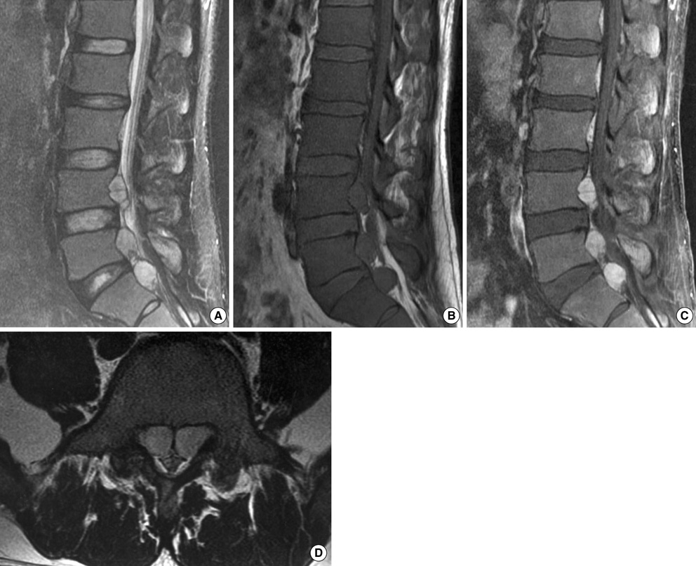
Fig. 1 Epidural extramedullary hematopoiesis in idiopathic myelofibrosis. Sagittal T2 (A), T1-weighted (B), and enhanced magnetic resonance (MR) images (C) of the lumbar spine revealing a posterior L4-S1 epidural mass causing spinal cord compression, as a homogenous isointense signal on T1-weighted and slightly hyperintense signal on T2-weighted images compared with adjacent red bone marrow, which were well enhanced with contrast (Gadolinium). Axial T2- weighted images (D) reveal an extradural mass extending into the neural foramen bilaterally on the L5 vertebral level.
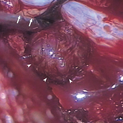
Fig. 2 Intraoperative photograph showing the discrete, flesh-colored, elastic, and extradural mass (arrowheads) between lumbar nerve roots (arrows).
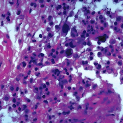
Fig. 3 Microscopic photograph of the extradural mass revealing cellular tissue of hematopoietic origin with small megakaryocytes (H&E, ×400).
Reference
-
1. Kwak HS, Lee JM. CT findings of extramedullary hematopoiesis in the thorax, liver and kidneys, in a patient with idiopathic myelofibrosis. J Korean Med Sci. 2000. 15:460–462.
Article2. Cook G, Sharp RA. Spinal cord compression due to extramedullary haemopoiesis in myelofibrosis. J Clin Pathol. 1994. 47:464–465.
Article3. De Klippel N, Dehou MF, Bourgain C, Schots R, De Keyser J, Ebinger G. Progressive paraparesis due to thoracic extramedullary hematopoiesis in myelofibrosis. Case report. J Neurosurg. 1993. 79:125–127.4. McDonald AC, Cook G, Sharp RA, Bissett D. Spinal cord compression in myelofibrosis--a case report. Acta Oncol. 1993. 32:692–693.
Article5. Guermazi A, Miaux Y, Chiras J. Imaging of spinal cord compression due to thoracic extramedullary haematopoiesis in myelofibrosis. Neuroradiology. 1997. 39:733–736.
Article6. de Haas KP, van de Loosdrecht AA, Daenen SM. Intraspinal extramedullary haematopoiesis in a patient with myelofibrosis. Neth J Med. 2002. 60:256–259.7. Horwood E, Dowson H, Gupta R, Kaczmarski R, Williamson M. Myelofibrosis presenting as spinal cord compression. J Clin Pathol. 2003. 56:154–156.
Article8. Close AS, Taira Y, Cleveland DA. Spinal cord compression due to extramedullary hematopoiesis. Ann Intern Med. 1958. 48:421–427.
Article9. Saghafi M, Shirdel A, Lari SM. Extramedullary hematopoiesis with spinal cord compression in beta-thalassemia intermedia. Eur J Intern Med. 2005. 16:596–597.10. Salehi SA, Koski T, Ondra SL. Spinal cord compression in beta-thalassemia: case report and review of the literature. Spinal Cord. 2004. 42:117–123.
Article11. Jalbert F, Chaynes P, Lagarrigue J. Asymptomatic spherocytosis presenting with spinal cord compression: case report. J Neurosurg Spine. 2005. 2:491–494.12. Shin KH, Sharma S, Gregoritch SJ, Lifeso RM, Bettigole R, Yoon SS. Combined radiotherapeutic and surgical management of a spinal cord compression by extramedullary hematopoiesis in a patient with hemoglobin E beta-thalassemia. Acta Haematol. 1994. 91:154–157.
Article13. Ohta Y, Shichinohe H, Nagashima K. Spinal cord compression due to extramedullary hematopoiesis associated with polycythemia vera--case report. Neurol Med Chir (Tokyo). 2002. 42:40–43.14. Niggemann P, Krings T, Hans F, Thron A. Fifteen-year follow-up of a patient with beta thalassaemia and extramedullary haematopoietic tissue compressing the spinal cord. Neuroradiology. 2005. 47:263–266.
Article
- Full Text Links
-
- Actions
-
Cited
- CITED
-
- Close
- Share
-
- Similar articles
-
- Cutaneous Extramedullary Hematopoiesis in Idiopathic Myelofibrosis
- A Case of Cutaneous Extramedullary Hematopoiesis in Idiopathic Myelofibrosis
- Two Cases of Myelofibrosis Mimicking Malignant Lymphoma in Computed Tomography of Abdomen: A Case of Autoimmune Myelofibrosis associated with Systemic Lupus Erythematosus Showing Extensive Lymphadenopathy and A Case of Chronic Idiopathic Myelofibrosis wit
- Cavernous Hemangioma of the Cauda Equina
- A Case of Chronic Myelogenous Leukemia with Extramedullary Hematopoiesis Presenting as Liver Mass
