Adenocarcinoma Arising in Gastric Heterotopic Pancreas: A Case Report
- Affiliations
-
- 1Department of Pathology, University of Ulsan College of Medicine, Asan Medical Center, Seoul, Korea. jskim@amc.seoul.kr
- 2Department of General Surgery, University of Ulsan College of Medicine, Asan Medical Center, Seoul, Korea.
- KMID: 1785711
- DOI: http://doi.org/10.3346/jkms.2004.19.1.145
Abstract
- A heterotopic pancreas in the gastrointestinal tract is mostly found incidentally and its malignant transformation is extremely rare. We describe the second case of adenocarcinoma arising in a gastric heterotopic pancreas of an asymptomatic 35-yr-old man in Korea. Esophagogastroduodenoscopy revealed a submucosal tumor with an irregular central umbilication in the gastric antrum. A wedge resection specimen demonstrated a submucosal oligolocular cystic mass (1.7x1.4x1.2 cm) with a solid portion. Microscopically, the cystic portion was composed of dilated pancreaticobiliary type ducts with adjacent small foci of periductal glandular structures. The adenocarcinoma components in the solid area infiltrated the proper muscle and the overlying mucosa of the stomach. The transitional area between the benign ductal structures and the adenocarcinoma component was found. The follow-up course was uneventful 5 months postoperatively.
Keyword
MeSH Terms
Figure
-

Fig. 1 The EGD shows a submucosal tumor with an irregular central umbilication in the gastric antrum.
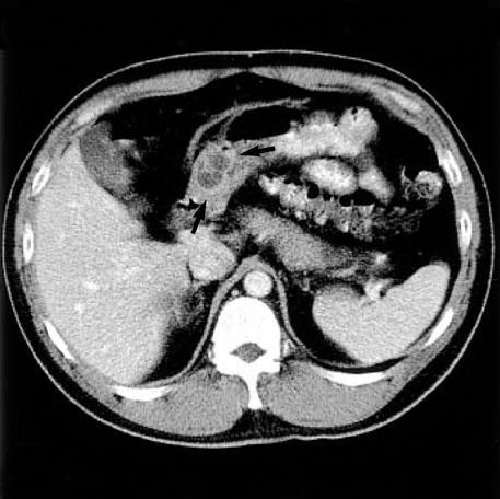
Fig. 2 The abdominal CT reveals a well-demarcated multiseptate cystic mass (arrows) in the gastric antrum.
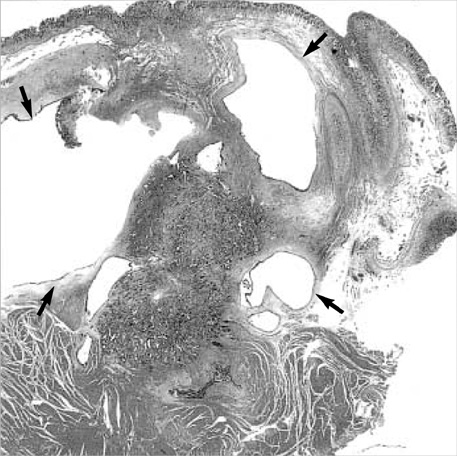
Fig. 3 The submucosal mass consists of oligolocular cystic (arrows) and solid areas without overlying mucosal change (H&E stain, ×10).

Fig. 4 The cystic area (arrows in Fig. 3) shows dilated benign ductal structures lined by cuboidal epithelium (A, H&E stain, ×200) and focal periductal glandular structures without islet cell islands (B, H&E stain, ×100).
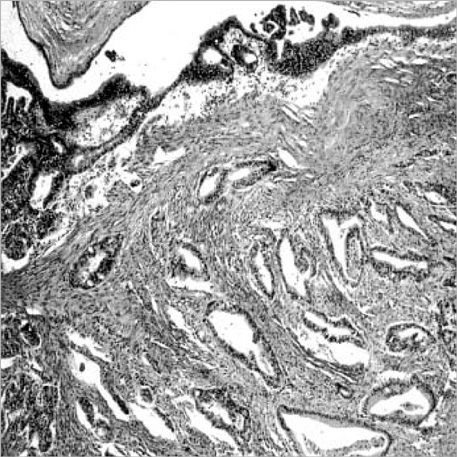
Fig. 5 The solid area shows well to moderately differentiated adenocarcinoma with an adjacent dysplastic change in the lining cells of the dilated ductal structures (H&E stain, ×40).
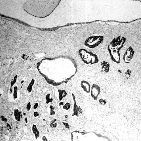
Fig. 6 Positive immunoreactivity for cytokeratin 7 is observed in the tumor cells and ductal epithelium in the heterotopic pancreas (×40).
Cited by 3 articles
-
첫 진단 10년 후 위출구폐색 증상으로 나타난 이소성 췌장에서 기인한 위선암
Hee Soo Jung, Jin Lee, Kyung Han Nam, Su Jin Jeong, Eun Hye Oh, Yong Eun Park, Jongha Park, Tae Oh Kim
Korean J Gastroenterol. 2020;76(1):37-41. doi: 10.4166/kjg.2020.76.1.37.Partial gastric outlet obstruction caused by a huge submucosal tumor originating in the heterotopic pancreas
Gum O Jung, Dong Eun Park, Ki Jung Yun, Kwon Mook Chae
Korean J Hepatobiliary Pancreat Surg. 2011;15(3):194-197. doi: 10.14701/kjhbps.2011.15.3.194.A Rare Case of Early Gastric Cancer Combined with Underlying Heterotopic Pancreas
Jung Bin Yoon, Bong Eun Lee, Dae Hwan Kim, Do Youn Park, Hye Kyung Jeon, Dong Hoon Baek, Gwang Ha Kim, Geun Am Song
Clin Endosc. 2018;51(2):192-195. doi: 10.5946/ce.2017.055.
Reference
-
1. Herold G, Kraft K. Adenocarcinoma arising from ectopic gastric pancreas: two case reports with a review of the literature. Z Gastroenterol. 1995. 33:260–264.2. St-Vil D, Brandt ML, Panic S, Bensoussan AL, Blanchard H. Meckel's diverticulum in children: a 20-year review. J Pediatr Surg. 1991. 26:1289–1292.
Article3. Qizilbash AH. Acute pancreatitis occurring in heterotopic pancreatic tissue in the gallbladder. Can J Surg. 1976. 19:413–414.4. Caberwal D, Kogan SJ, Levitt SB. Ectopic pancreas presenting as an umbilical mass. J Pediatr Surg. 1977. 12:593–599.
Article5. Ishikawa O, Ishiguro S, Ohhigashi H, Sasaki Y, Yasuda T, Imaoka S, Iwanaga T, Nakaizumi A, Fujita M, Wada A. Solid and papillary neoplasm arising from an ectopic pancreas in the mesocolon. Am J Gastroenterol. 1991. 85:597–601.6. Kakizaki Y, Ishiwatari Z, Hukushi K. A case of submucosal cyst of the stomach originating in the heterotopic pancreas. Stomach Intestine. 1975. 10:1651–1657.7. Kaneda M, Yano T, Yamamoto T, Suzuki T, Fujimori K, Itoh H, Mizumoto R. Ectopic pancreas in the stomach presenting as an inflammatory abdominal mass. Am J Gastroenterol. 1989. 84:663–666.8. Makhlouf HR, Almeida JL, Sobin LH. Carcinoma in jejunal pancreatic heterotopia. Arch Pathol Lab Med. 1999. 123:707–711.
Article9. Jeng KS, Yang KC, Kuo SHF. Malignant degeneration of heterotopic pancreas. Gastrointest Endosc. 1991. 37:196–198.
Article10. Ahn YS, Cho JS, Shin KS, Noh SM, Jeong HY, Song KS. Ductal adenocarcinoma arising from heterotopic pancreas in the stomach; a case report. J Korean Radiol Soc. 2001. 45:51–53.
Article11. Chapple CR, Muller S, Newman J. Gastric adenocarcinoma associated with adenomyoma of the stomach. Postgrad Med J. 1998. 64:801–803.
Article12. Kneafsey PD, Demetrick DJ. Malignant transformation in a pyloric adenomyoma: a case report. Histopathology. 1992. 20:433–435.
Article13. Osanai M, Miyokawa N, Tamaki T, Yonekawa M, Kawamura A, Sawada N. Adenocarcinoma arising in gastric heterotopic pancreas: clinicopathological and immunohistochemical study with genetic analysis of a case. Pathol Int. 2001. 51:549–554.
Article14. Duval JV, Savas L, Banner BF. Expression of cytokeratin 7 and 20 in carcinomas of the extrahepatic biliary tract, pancreas, and gallbladder. Arch Pathol Lab Med. 2000. 124:1196–1200.15. von Heinrich H. Ein Beitrag zur Histologie des sogen: Akzessorischen Pankreas. Virchows Arch A Pathol Anat Histopathol. 1909. 198:392–401.16. Hickman DM, Frey CF, Carson JW. Adenocarcinoma arising in gastric heterotopic pancreas. West J Med. 1981. 135:57–62.
- Full Text Links
-
- Actions
-
Cited
- CITED
-
- Close
- Share
-
- Similar articles
-
- Ductal Adenocarcinoma Arising from Heterotopic Pancreas in the Stomach: A Case Report
- Gastric Adenocarcinoma Arising from Heterotopic Pancreas Presenting as Gastric Outlet Obstruction 10 Years after the First Diagnosis
- A Case of Duodenal Adenocarcinoma Arising from the Heterotopic Pancreas
- Ductal Adenocarcinoma Arising from the Heterotopic Pancreas Situated in the Jejunum
- Colonic Adenocarcinoma Arising from Gastric Heterotopia: A Case Study
