Korean J Radiol.
2010 Apr;11(2):141-148. 10.3348/kjr.2010.11.2.141.
Histopathologic Findings Related to the Indeterminate or Inadequate Results of Fine-Needle Aspiration Biopsy and Correlation with Ultrasonographic Findings in Papillary Thyroid Carcinomas
- Affiliations
-
- 1Department of Radiology, Seoul St. Marys Hospital, The Catholic University of Korea, Seoul 137-701, Korea. skchung@catholic.ac.kr
- 2Department of Pathology, Seoul St. Marys Hospital, The Catholic University of Korea, Seoul 137-701, Korea.
- 3Department of Endoclinology, Seoul St. Marys Hospital, The Catholic University of Korea, Seoul 137-701, Korea.
- 4Department of Surgery, Seoul St. Marys Hospital, The Catholic University of Korea, Seoul 137-701, Korea.
- KMID: 1783189
- DOI: http://doi.org/10.3348/kjr.2010.11.2.141
Abstract
OBJECTIVE
To determine histopathologic findings related to the indeterminate or inadequate result of fine-needle aspiration biopsy (FNAB) in papillary thyroid carcinomas (PTCs) and to correlate histopathological findings with ultrasonographic features of tumors. Materials and
METHODS
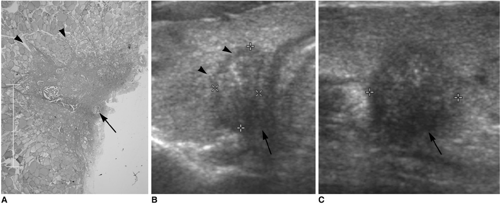
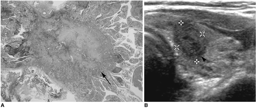
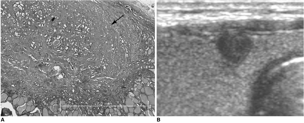
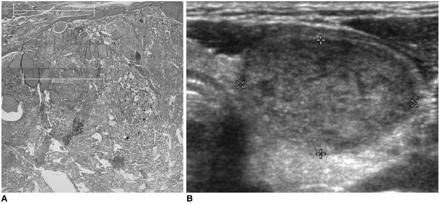
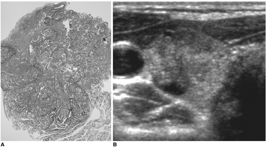
We retrospectively reviewed the medical records of FNAB, histopathologic characteristics, and sonographic findings of the solid portion of 95 PTCs in 95 patients. All cases were pathologically confirmed by surgery. Histopathologic characteristics were analyzed for tumor distribution, microcystic changes, fibrosis, and tumor component. We assumed several histopathologic conditions to be the cause of indeterminate or inadequate results of FNAB, including: 1) an uneven tumor distribution, 2) > 30% microcystic changes, 3) > 30% fibrosis, and 4) < 30% tumor component. Ultrasonographic findings of each PTC were evaluated for echotexture (homogeneous or heterogeneous), echogenicity (markedly hypoechoic, hypoechoic, isoechoic, or hyperechoic), and volume of the nodule. We correlated histopathologic characteristics of the PTC with results of the FNAB and ultrasonographic findings.
RESULTS
From 95 FNABs, 71 cases (74%) were confirmed with malignancy or suspicious malignancy (PTCs), 21 (22%) had indeterminate results (atypical cells), and three (4%) were negative for malignancy. None of the assumed variables influenced the diagnostic accuracy of FNAB. Tumor distribution and fibrosis were statistically correlated with ultrasonographic findings of the PTCs (p < 0.05). Uneven tumor distribution was related with small tumor volume, and fibrosis over 30% was correlated with homogeneous echotexture, markedly hypoechoic and hypoechoic echogenicity, and small tumor volume (p < 0.05).
CONCLUSION
No histopathologic component was found to correlate with improper results of FNAB in PTCs. In contrast, two histopathologic characteristics, uneven distribution and fibrosis, were correlated with ultrasonographic findings.
Keyword
MeSH Terms
Figure
Cited by 2 articles
-
Ultrasonographic Echogenicity and Histopathologic Correlation of Thyroid Nodules in Core Needle Biopsy Specimens
Ji-hoon Kim, Dong Gyu Na, Hunkyung Lee
Korean J Radiol. 2018;19(4):673-681. doi: 10.3348/kjr.2018.19.4.673.Degenerating Thyroid Nodules: Ultrasound Diagnosis, Clinical Significance, and Management
Jie Ren, Jung Hwan Baek, Sae Rom Chung, Young Jun Choi, Chan Kwon Jung, Jeong Hyun Lee
Korean J Radiol. 2019;20(6):947-955. doi: 10.3348/kjr.2018.0599.
Reference
-
1. Alexander EK, Heering JP, Benson CB, Frates MC, Doubilet PM, Cibas ES, et al. Assessment of nondiagnostic ultrasound-guided fine needle aspirations of thyroid nodules. J Clin Endocrinol Metab. 2002. 87:4924–4927.2. Danese D, Sciacchitano S, Farsetti A, Andreoli M, Pontecorvi A. Diagnostic accuracy of conventional versus sonography-guided fine-needle aspiration biopsy of thyroid nodules. Thyroid. 1998. 8:15–21.3. Rosen IB, Azadian A, Walfish PG, Salem S, Lansdown E, Bedard YC. Ultrasound-guided fine-needle aspiration biopsy in the management of thyroid disease. Am J Surg. 1993. 166:346–349.4. Chan BK, Desser TS, McDougall IR, Weigel RJ, Jeffrey RB Jr. Common and uncommon sonographic features of papillary thyroid carcinoma. J Ultrasound Med. 2003. 22:1083–1090.5. Moon WJ, Jung SL, Lee JH, Na DG, Baek JH, Lee YH, et al. Benign and malignant thyroid nodules: US differentiation--multicenter retrospective study. Radiology. 2008. 247:762–770.6. Kim EK, Park CS, Chung WY, Oh KK, Kim DI, Lee JT, et al. New sonographic criteria for recommending fine-needle aspiration biopsy of nonpalpable solid nodules of the thyroid. AJR Am J Roentgenol. 2002. 178:687–691.7. Chen SJ, Yu SN, Tzeng JE, Chen YT, Chang KY, Cheng KS, et al. Characterization of the major histopathological components of thyroid nodules using sonographic textural features for clinical diagnosis and management. Ultrasound Med Biol. 2009. 35:201–208.8. McHenry CR, Walfish PG, Rosen IB. Non-diagnostic fine needle aspiration biopsy: a dilemma in management of nodular thyroid disease. Am Surg. 1993. 59:415–419.9. Chow LS, Gharib H, Goellner JR, van Heerden JA. Nondiagnostic thyroid fine-needle aspiration cytology: management dilemmas. Thyroid. 2001. 11:1147–1151.10. Carmeci C, Jeffrey RB, McDougall IR, Nowels KW, Weigel RJ. Ultrasound-guided fine-needle aspiration biopsy of thyroid masses. Thyroid. 1998. 8:283–289.11. Kim DW, Park AW, Lee EJ, Choo HJ, Kim SH, Lee SH, et al. Ultrasound-guided fine-needle aspiration biopsy of thyroid nodules smaller than 5 mm in the maximum diameter: assessment of efficacy and pathological findings. Korean J Radiol. 2009. 10:435–440.12. Kim DW, Lee EJ, Kim SH, Kim TH, Lee SH, Kim DH, et al. Ultrasound-guided fine-needle aspiration biopsy of thyroid nodules: comparison in efficacy according to nodule size. Thyroid. 2009. 19:27–31.13. Livolsi VA. Papillary neoplasms of the thyroid. Pathologic and prognostic features. Am J Clin Pathol. 1992. 97:426–434.14. Isarangkul W. Dense fibrosis. Another diagnostic criterion for papillary thyroid carcinoma. Arch Pathol Lab Med. 1993. 117:645–646.15. Di Pasquale M, Rothstein JL, Palazzo JP. Pathologic features of Hashimoto's-associated papillary thyroid carcinomas. Hum Pathol. 2001. 32:24–30.
- Full Text Links
- Actions
-
Cited
- CITED
-
- Close
- Share
- Similar articles
-
- Evaluating the Degree of Conformity of Papillary Carcinoma and Follicular Carcinoma to the Reported Ultrasonographic Findings of Malignant Thyroid Tumor
- Thyroid nodules with discordant results of ultrasonographic and fine-needle aspiration findings
- Oxyphilic Papillary Carcinoma of the Thyroid in Fine Needle Aspiration
- Current Guidelines for Fine Needle Aspiration of Thyroid Nodules
- Fine Needle Aspiration Cytology of Columnar Cell Variant of Papillary Carcinoma of the Thyroid: A Case Report

