Korean J Radiol.
2005 Mar;6(1):37-40. 10.3348/kjr.2005.6.1.37.
Posttraumatic Pseudoaneurysm in Scalp Treated by Direct Puncture Embolization Using N-Butyl-2-Cyanoacrylate: a Case Report
- Affiliations
-
- 1Department of Neurosurgery, Seoul National University Boramae Hospital, Korea.
- 2Department of Radiology, Seoul National University Boramae Hospital, Korea. cyho50168@radiol.snu.ac.kr
- KMID: 1783171
- DOI: http://doi.org/10.3348/kjr.2005.6.1.37
Abstract
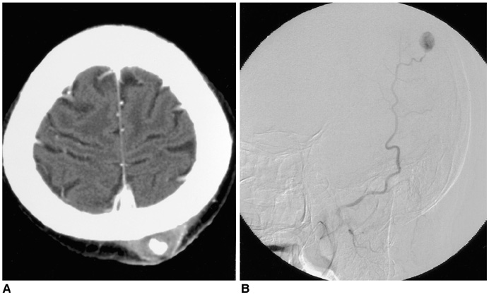
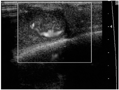
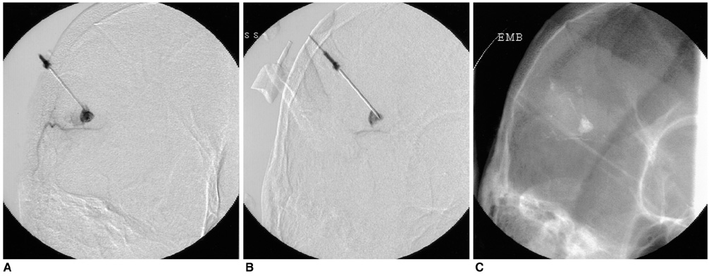
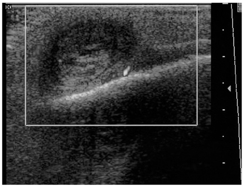
- Here, we report a case of scalp pseudoaneurysm which was treated by direct puncture embolization using n-butyl-2-cyanoacrylate. The patient had a history of blunt trauma in the previous two months. Ultrasound-guided manual compression was initially attempted, but the results were unsatisfactory. Direct puncture embolization was then performed, and the pseudoaneurysm was completely obliterated. Non-surgical treatment options for pseudoaneurysm are briefly discussed.
Keyword
MeSH Terms
Figure
Reference
-
1. Kang SS, Labropoulos N, Mansour MA, Baker WH. Percutaneous ultrasound guided thrombin injection: a new method for treating postcatheterization femoral pseudoaneurysms. J Vasc Surg. 1998. 27:1032–1038.2. Paulson EK, Sheafor DH, Kliewer MA, Nelson RC, Eisenberg LB, Sebastian MW, et al. Treatment of iatrogenic femoral arterial pseudoaneurysms: comparison of US-guided thrombin injection with compression repair. Radiology. 2000. 215:403–408.3. Isaacson G, Kochan PS, Kochan JP. Pseudoaneurysms of the superficial temporal artery: treatment options. Laryngoscope. 2004. 114:1000–1004.4. Conner WC 3rd, Rohrich RJ, Pollock RA. Traumatic aneurysms of the face and temple: a patient report and literature review, 1644 to 1998. Ann Plast Surg. 1998. 41:321–326.5. Cadamy AJ, McNaughton GW, Helliwell R. Traumatic pseudoaneurysms of the superficial temporal artery. Eur J Emerg Med. 2003. 10:236–237.6. Paulson EK, Kliewer MA, Hertzberg BS, Tcheng JE, McCann RL, Bowie JD, et al. Ultrasonographically guided manual compression of femoral artery injuries. J Ultrasound Med. 1995. 14:653–659.7. Partap VA, Cassoff J, Glikstein R. US-guided percutaneous thrombin injection: a new method of repair of superficial temporal artery pseudoaneurysm. J Vasc Interv Radiol. 2000. 11:461–463.8. Teh LG, Sieunarine K. Thrombin injection for repair of pseudoaneurysms: a case for caution. Australas Radiol. 2003. 47:64–66.9. Choi YH, Han MH, O-Ki K, Cha SH, Chang KH. Craniofacial cavernous venous malformations: percutaneous sclerotherapy with use of ethanolamine oleate. J Vasc Interv Radiol. 2002. 13:475–482.
- Full Text Links
- Actions
-
Cited
- CITED
-
- Close
- Share
- Similar articles
-
- Percutaneous N-Butyl Cyanoacrylate Embolization of a Pancreatic Pseudoaneurysm after Failed Attempts of Transcatheter Embolization
- Multiple Cerebral Infarction after Injection of N-Butyl-2-Cyanoacrylate for Gastric Variceal Bleeding
- Transcatheter Arterial Embolization of Cystic Artery Pseudoaneurysm in Acalculous Cholecystitis: A Case Report
- Transcatheter Arterial Embolization with N-Butyl Cyanoacrylate for Profunda Femoris Artery Pseudoaneurysms Caused by Femur Shaft Fractures: Two Case Reports
- Direct Puncture Embolization of Scalp Arteriovenous Malformation in a Patient with Severe Hemophilia A: A Case Report


