Lymphobronchial Fistula of Tuberculous Lymphadenitis in Acquired Immunodeficiency Syndrome
- Affiliations
-
- 1Department of Internal Medicine, National Medical Center, Seoul, Korea. ssabana777@gmail.com
- KMID: 1782397
- DOI: http://doi.org/10.3947/ic.2012.44.1.35
Abstract
- Bronchial invasion of tuberculous lymphadenitis in children has been reported in areas of high tuberculosis (TB) prevalence as a complication due to primary pulmonary tuberculosis. However, it is rare in immunocompetent adults. When it appears, it often presents as a consequence of the reactivation of TB in the lung parenchyma. Primary TB occurs more frequently in patients with human immunodeficiency virus (HIV), with a history of organ transplants, or undergoing immunosuppressive treatments such as steroids. Furthermore, bronchial invasion of the bronchus by tuberculous lymphadenitis is considered to be very rare even among immunocompromised adults with primary TB, and has never before been reported in Korea. The authors report a case of bronchial invasion of the bronchus by tuberculous lymphadenitis, confirmed by bronchoscopy, in an Acquired Immunodeficiency Syndrome (AIDS) patient.
Keyword
MeSH Terms
Figure
-
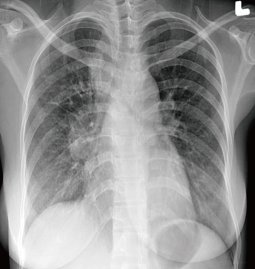
Figure 1 Chest radiography shows mediastinal widening and right hilar bulging without parenchymal infiltration on admission.
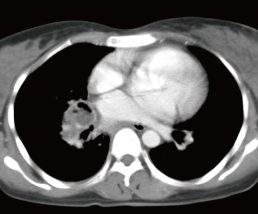
Figure 2 Chest CT scan shows the enlargement of peribronchial lymph node in the right middle lobe bronchus.
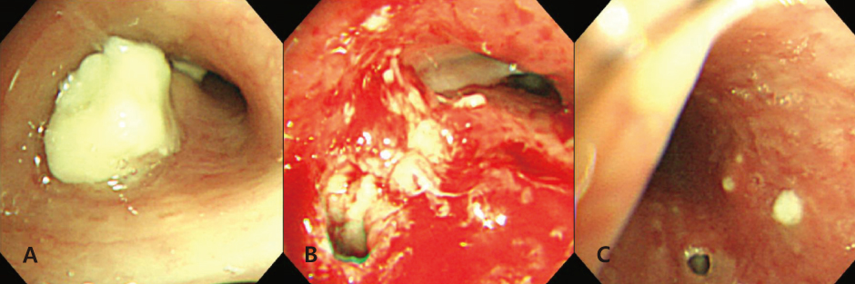
Figure 3 Bronchoscopic findings: (A) Right middle lobe bronchus is filled with white caseous material, which is in agreement with the soft tissue abnormality seen on the chest CT. (B) A fistula was discovered after the removal of the white caseous material. (C) A fistula and white node-like spots are shown on the left main bronchus. These bronchoscopic findings suggested the infiltration of the TB lymphadenitis into the bronchus.

Figure 4 Histopathologic finding of bronchoscopic biopsy: (A) Caseous necrosis and lymphocyte infiltration (H&E stain, ×400). (B) Positive acid fast bacilli (AFB) are present (AFB stain, ×1,000).
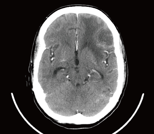
Figure 5 Contrast-enhanced brain CT. A mass with edema was shown on the left frontal area surrounded by a hyperdense lesion.

Figure 6 T1 weighted brain MRI shows multiple enhanced lesions.
Cited by 1 articles
-
Polymicrobial Purulent Pericarditis Probably caused by a Broncho-Lymph Node-Pericardial Fistula in a Patient with Tuberculous Lymphadenitis
Seung Lee, Kanglok Lee, Jun Kwon Ko, Jaekeun Park, Mi Yeon Yu, Chang Kyo Oh, Seung Pyo Hong, Yeonjae Kim, Younghyo Lim, Hyuck Kim, Hyunjoo Pai
Infect Chemother. 2015;47(4):261-267. doi: 10.3947/ic.2015.47.4.261.
Reference
-
1. Auerbach O. Tuberculosis of the trachea and major bronchi. Am Rev Tuberc. 1949. 60:604–620.2. Wasser LS, Shaw GW, Talavera W. Endobronchial tuberculosis in the acquired immunodeficiency syndrome. Chest. 1988. 94:1240–1244.
Article3. Goussard P, Gie R. Airway involvement in pulmonary tuberculosis. Paediatr Respir Rev. 2007. 8:118–123.
Article4. FitzGerald JM, Grzybowski S, Allen EA. The impact of human immunodeficiency virus infection on tuberculosis and its control. Chest. 1991. 100:191–200.
Article5. Leung AN, Brauner MW, Gamsu G, Mlika-Cabanne N, Ben Romdhane H, Carette MF, Grenier P. Pulmonary tuberculosis: comparison of CT findings in HIV-seropositive and HIV-seronegative patients. Radiology. 1996. 198:687–691.
Article6. Daley CL, Small PM, Schecter GF, Schoolnik GK, McAdam RA, Jacobs WR Jr, Hopewell PC. An outbreak of tuberculosis with accelerated progression among persons infected with the human immunodeficiency virus. An analysis using restriction-fragment-length polymorphisms. N Engl J Med. 1992. 326:231–235.
Article7. Golden MP, Vikram HR. Extrapulmonary tuberculosis: an overview. Am Fam Physician. 2005. 72:1761–1768.8. Pitchenik AE, Rubinson HA. The radiographic appearance of tuberculosis in patients with the acquired immune deficiency syndrome (AIDS) and pre-AIDS. Am Rev Respir Dis. 1985. 131:393–396.9. Judson MA, Sahn SA. Endobronchial lesions in HIV-infected individuals. Chest. 1994. 105:1314–1323.
Article10. Smart J. Endobronchial tubercuosis. Br J Dis Chest. 1951. 45:61–68.11. Kashyap S, Mohapatra PR, Saini V. Endobronchial tuberculosis. Indian J Chest Dis Allied Sci. 2003. 45:247–256.12. Freixinet J, Varela A, Lopez Rivero L, Caminero JA, Rodríguez de Castro F, Serrano A. Surgical treatment of childhood mediastinal tuberculous lymphadenitis. Ann Thorac Surg. 1995. 59:644–646.
Article13. Frostad S. Segmental atelectasis in children with primary tuberculosis. Am Rev Tuberc. 1959. 79:597–605.
Article
- Full Text Links
-
- Actions
-
Cited
- CITED
-
- Close
- Share
-
- Similar articles
-
- A Case of Urethral Diverticulo-Rectal Fistula in Acquired Immunodeficiency Syndrome
- AIDS Diagnosed in the Course of Managing Duodenal Fistula Caused by Tuberculosis: A Case Report
- Esophago-Mediastinal Fistula Due to Tuberculous Mediastinal Lymphadenitis
- A Case of Nontuberculous Mycobacterium Infection Complicated by an Esophagomediastinal Fistula in a Human Immunodeficiency Virus Patient
- Cutaneous Cytomegalovirus Infection Presenting as Papules and Pustules in a Patient with Acquired Immunodeficiency Syndrome
