A Case of Klebsiella pneumoniae Liver Abscess Which Progressed to Pyomyositis and Infected Aneurysm of Aorta
- Affiliations
-
- 1Department of Internal Medicine, Division of Infectious Disease, Hanyang University College of Medicine, Seoul, Korea. paihj@hanyang.ac.kr
- KMID: 1782303
- DOI: http://doi.org/10.3947/ic.2008.40.6.341
Abstract
- Klebsiella pneumoniae causes pyogenic infections in various sites, with the risk of which increases in patients with diabetes mellitus. Recently, K. pneumoniae has emerged as a leading cause of pyogenic liver abscess. Primary liver abscess caused by K. pneumoniae in the absence of underlying hepatobiliary disease is commonly associated with metastatic infections such as endophthalmitis, meningitis, brain abscess and infection in other sites. We experienced a case of K. pneumoniae liver abscess associated with septic metastatic lesions including pyomyositis and infected aneurysm of aorta. Despite the aggressive management with antibiotics, surgical pus drainage and aortic bypass graft, patient died of ventilator associated pneumonia and multiorgan failure. Our experience suggests that clinicians should be alert to septic metastatic infections when they treat a patient with K. pneumoniae liver abscess.
MeSH Terms
Figure
-
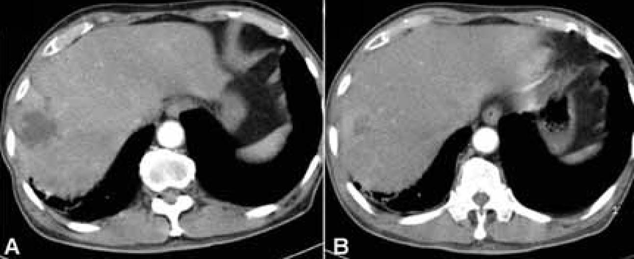
Figure 1 (A) Abdominal CT scan reveals about 3 cm sized lobulated liver abscess in segment 8. (B) Follow up abdominal CT scan after two months shows decreased size of liver abscess.
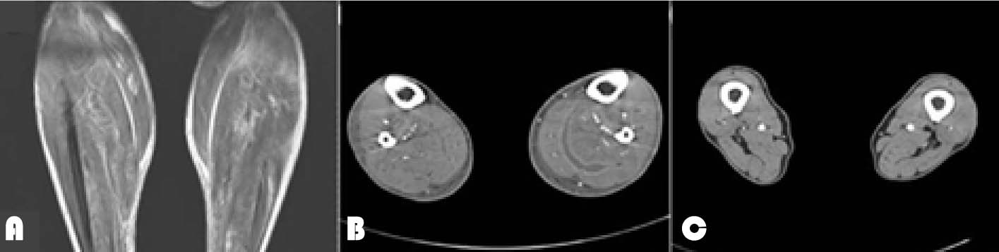
Figure 2 (A) Coronal STIR MR image of both calves on admission shows high signal intensity along the calf muscle, fascia, and subcutaneous tissue. (B) CT scan of lower extremities on the 9th hospital day shows extensive fluid collections with peripheral enhancement within the muscle of both lower legs. (C) Follow up CT scan on the 23th hospital day shows regression of the abscess.
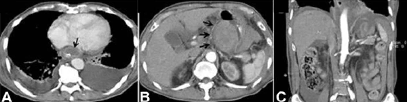
Figure 3 Abdominal CT scan on the 32th hospital day shows a well localized hematoma involving the distal esophagus (arrow) (A) which extends across the gastroesophageal junction into the body of the stomach (arrow) (B). High-attenuation fluid in perigastric, perisplenic and perihepatic area is noted and suggests hemoperitoneum (C).
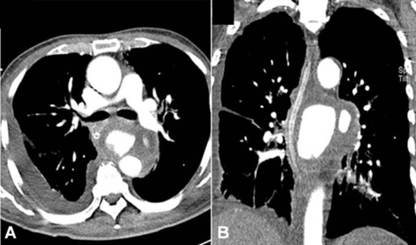
Figure 4 Chest CT scan shows proximal descending aortic aneurysm measuring 9 cm in diameter with diffuse hematoma in paraesophageal area.
Reference
-
1. Braiteh F, Golden MP. Cryptogenic invasive Klebsiella pneumoniae liver abscess syndrome. Int J Infect Dis. 2007. 11:16–22.2. Ko WC, Paterson DL, Sagnimeni AJ, Hansen DS, Von Gottberg A, Mohapatra S, Casellas JM, Goossens H, Mulazimoglu L, Trenholme G, Klugman KP, McCormack JG, Yu VL. Community-acquired Klebsiella pneumoniaeiae bacteremia: global differences in clinical patterns. Emerg Infect Dis. 2002. 8:160–166.
Article3. Park SH, Choi SM, Nam KW, Kim SI, Wie SH, Kim YR, Moon IS, Kang MW. Two cases of Klebsiella pneumoniae liver abscess complicated with multiple septic metastatic lesions: their association with diabetes mellitus. Korean J Infect Dis. 2001. 33:364–370.4. Chung DR, Lee SS, Lee HR, Kim HB, Choi HJ, Eom JS, Kim JS, Choi YH, Lee JS, Chung MH, Kim YS, Lee H, Lee MS, Park CK. Korean Study Group for Liver Abscess Emerging invasive liver abscess caused by K1 serotype Klebsiella pneumoniae in Korea. J Infect. 2007. 54:578–583.
Article5. Wang JH, Liu YC, Lee SS, Yen MY, Chen YS, Wang JH, Wann SR, Lin HH. Primary liver abscess due to Klebsiella pneumoniae in Taiwan. Clin Infect Dis. 1998. 26:1434–1438.
Article6. Woo SI, Kwon HC, Choi YH, Shin SS, Kim CO, Kim HY, Park YS, Park YS, Song YG, Yeom JS, Yoon HJ, Lee KS, Choi SH, Choi JY, Hong SK, Kim JM. Clinical characteristics of pyogenic liver abscess with Klebsiella pneumoniae or non-Klebsiella pneumoniae and its prognosis associated with diabetes mellitus. Infect Chemother. 2006. 38:77–84.7. Kim YS, Gang YS, Jung JH, Park KO, Kim SM, Lee BS, Lee HY. Septic metastatic bilateral endophthalmitis complicating Klebsiella pneumoniae liver abscess in a non-diabetic woman. Korean J Med. 2005. 68:233–237.8. Kim GS, Lee JH, Choi SA, Lim SR. A case of Klebsiella pneumoniae liver abscess complicated with brain abscess and endophthalmitis. J Korean Neurol Assoc. 2005. 23:578–580.9. Lee JY, Nah BK, Kim JH, Lee SH, Choi TH, Choi HY, Kim KS, Cheon GJ. Septic metastatic lesions associated with Klebsiella pneumoniae liver abscess. Infect Chemother. 2006. 38:95–100.10. Rogers DW, Lumeng L, Goulet RJ, Canal DF. Ruptured mycotic pseudoaneurysm of the gastroduodenal artery presenting with hemoperitoneum and subcapsular liver hematoma. Dig Dis Sci. 1990. 35:661–664.
Article11. Hu BS, Lau YJ, Shi ZY, Lin YH. Necrotizing fasciitis associated with Klebsiella pneumoniae liver abscess. Clin Infect Dis. 1999. 29:1360–1361.
Article12. Mincheff TV, Cooler AW. Ruptured mycotic aneurysm presenting initially with bacterial meningitis. Am Surg. 2008. 74:73–75.
Article13. Kim SG, Park DK, Koo YS, Kang DH, Choi DJ, Park HC, Kim JH, Son JW, Shin IK. A case of infected abdominal aortic aneurysm associated with liver abscess treated by endovascular stent. Korean J Med. 2001. 61:151–155.14. Sugawa M, Tanaka R, Nakamura M, Isaka N, Nishimura J, Kimura M, Nakano T. A case of infectious pseudoaneurysm of the abdominal aorta associated with infectious spondylitis due to Klebsiella pneumoniae. Jpn J Med. 1989. 28:402–405.
Article15. Sugawara Y, Sato O, Miyata T, Kimura H, Yamaoka M, Uozaki H, Oka T, Makuuchi M. Ruptured abdominal aorta secondary to psoas muscle abscess due to Klebsiella pneumoniae in an alcoholic. J Infect. 1997. 35:185–188.
Article
- Full Text Links
-
- Actions
-
Cited
- CITED
-
- Close
- Share
-
- Similar articles
-
- A Case of Liver Abscess Metastasizing to Prostate Resulting in Prostatic Abscess
- Metastatic endophthalmitis and thyroid abscess complicating Klebsiella pneumoniae liver abscess
- A case of infected abdominal aortic aneurysm associated with liver abscess treated by endovascular stent
- Liver abscess due to Klebsiella pneumoniae in a healthy 12-year-old boy
- Klebsiella pneumoniae Brain Abscess and Endophthalmitis after Acute Epiglottitis
