Korean J Radiol.
2006 Jun;7(2):80-86. 10.3348/kjr.2006.7.2.80.
CT Findings of Atypical Adenomatous Hyperplasia in the Lung
- Affiliations
-
- 1Department of Radiology, Seoul National University College of Medicine, and the Institute of Radiation Medicine, Seoul National University Medical Research Center, and the Clinical Research Institute, Seoul National University Hospital, Seoul, Korea. jmgo
- 2Department of Pathology, Seoul National University College of Medicine, Seoul, Korea.
- KMID: 1782180
- DOI: http://doi.org/10.3348/kjr.2006.7.2.80
Abstract
OBJECTIVE
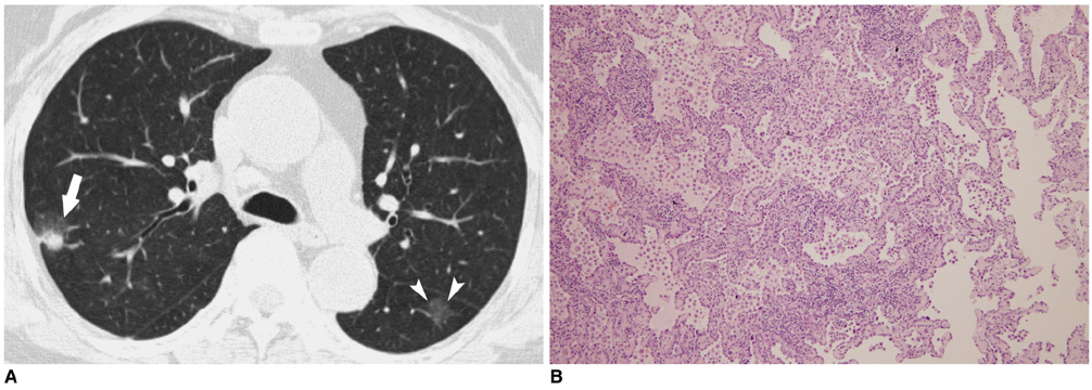
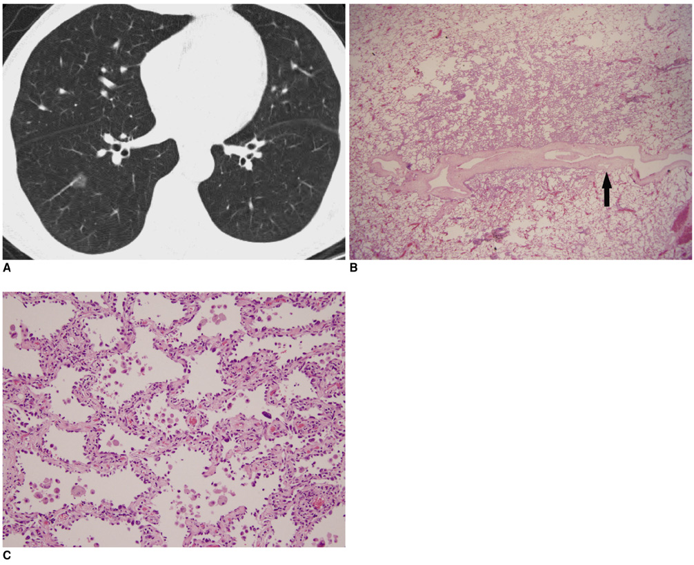
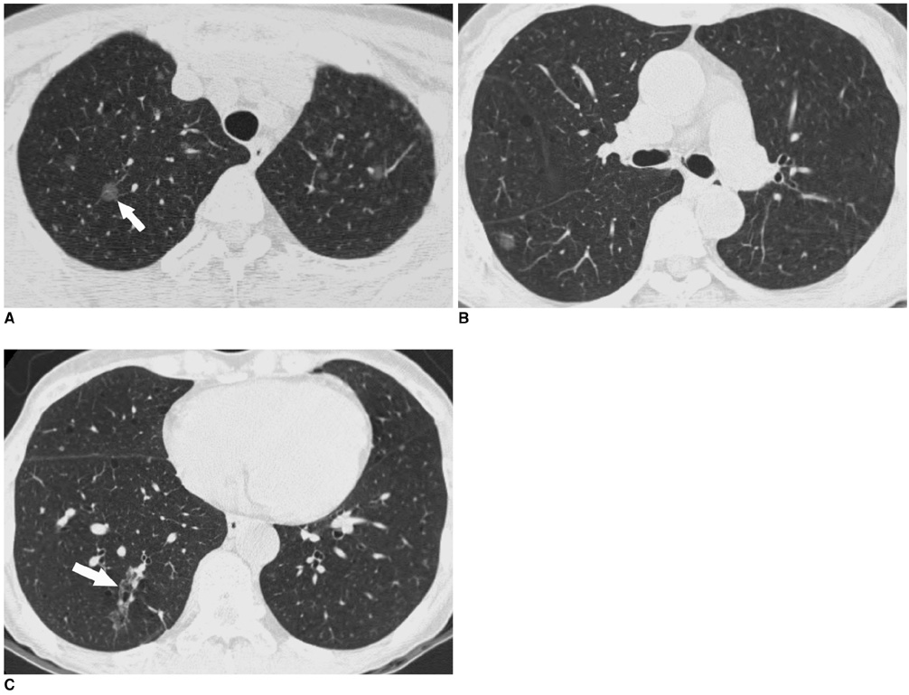
The aim of this study was to analyze the computed tomographic (CT) findings of atypical adenomatous hyperplasia (AAH) in the lung. MATERIALS AND METHODS: The CT findings of AAHs in eight patients were retrospectively reviewed. The CT findings of each AAH lesion were evaluated for multiplicity, location, shape, size and internal density of the lesion, the interface between the normal lung and the lesion, the internal features within the lesion and any change of the lesion on the follow-up CT scans (range: 33 to 540 days; average: 145.3 days). RESULTS: The eight patients consisted of three men and five women (age range: 43-71 years). Six of eight patients were asymptomatic. Four of them (50%) had synchronous malignancies in the lung: adenocarcinoma of the lung (n = 3), and metastatic squamous cell carcinoma from the uterus (n = 1). We could identify and evaluate eleven AAH nodules in seven patients on the CT scans. Three patients had multiple AAHs. Seven of the 11 lesions (64%) were located in the upper lobe. All the AAHs showed a well-defined oval or round shape and pure ground-glass opacity (GGO) without any solid component (size: 3.9x3 mm to 19x17 mm; internal attenuation: -467 to -785 HU). All the AAHs showed no change of their size and internal density on the follow-up CT scans. CONCLUSION: Atypical adenomatous hyperplasia is often associated with malignancy. This tumor is shown on CT as persistent well-defined oval or round nodular GGOs without solid components, and it does not change on the follow-up CT.
MeSH Terms
Figure
Cited by 1 articles
-
A Case of Atypical Adenomatous Hyperplasia of Larger Than 2 cm
Bo Mi Park, Min Ji Cho, Hyun Seok Lee, Dong Il Park, Myoung Rin Park, Ju Ock Kim, Jeong Eun Lee, Choong Sik Lee, Sung Soo Jung
Tuberc Respir Dis. 2013;74(6):280-285. doi: 10.4046/trd.2013.74.6.280.
Reference
-
1. Weng S, Tsuchiya E, Satoh Y, Kitagawa T, Nakagawa K, Sugano H. Multiple atypical hyperplasia of type II pneumocytes and bronchioloalveolar carcinoma. Histopathology. 1990. 16:101–103.2. Carey FA, Wallace WA, Fergusson RJ, Kerr KM, Lamb D. Alveolar atypical hyperplasia in association with primary pulmonary adenocarcinoma: a clinicopathological study of 10 cases. Thorax. 1992. 47:1041–1043.3. Colby TV, Wistuba II, Gazdar A. Precursors to pulmonary neoplasia. Adv Anat Pathol. 1998. 5:205–215.4. Yokozaki M, Kodama T, Yokose T, Matsumoto T, Mukai K. Differentiation of atypical adenomatous hyperplasia and adenocarcinoma of the lung by use of DNA ploidy and morphometric analysis. Mod Pathol. 1996. 9:1156–1164.5. Pueblitz S, Hieger LR. Expression of p53 and CEA in atypical adenomatous hyperplasia of the lung [letter]. Am J Surg Pathol. 1997. 21:867–868.6. Kitamura H, Kameda Y, Ito T, Hayashi H. Atypical adenomatous hyperplasia of the lung. Implications for the pathogenesis of peripheral lung adenocarcinoma. Am J Clin Pathol. 1999. 111:610–622.7. Kushihashi T, Munechika H, Ri K, Kubota H, Ukisu R, Satoh S, et al. Bronchioalveolar adenoma of the lung: CT-pathologic correlation. Radiology. 1994. 193:789–793.8. Sone S, Takashima S, Li F, Yang Z, Honda T, Maruyama Y, et al. Mass screening for lung cancer with mobile spiral computed tomography scanner. Lancet. 1998. 351:1242–1245.9. Austin JH, Muller NL, Friedman PJ, Hansell DM, Naidich DP, Remy-Jardin M, et al. Glossary of terms for CT of the lung: recommendations of the Nomenclature Committee of the Fleischner Society. Radiology. 1996. 200:327–331.10. Travis WD, Brambilla E, Juller-Hermelink HK, Harris CC. Pathology and genetics of tumours of the lung, pleura, thymus and heart. 2004. 1st ed. Lyon: IARC Press;73–75.11. Kaneko M, Eguchi K, Ohmatsu H, Kakinuma R, Naruke T, Suemasu K, et al. Peripheral lung cancer: screening and detection with low-dose spiral CT versus radiography. Radiology. 1996. 201:798–802.12. Henschke CI, McCauley DI, Yankelevitz DF, Naidich DP, McGuinness G, Miettinen OS, et al. Early lung cancer action project: overall design and findings from baseline screening. Lancet. 1999. 354:99–105.13. Yokose T, Doi M, Tanno K, Yamazaki K, Ochiai A. Atypical adenomatous hyperplasia of the lung in autopsy cases. Lung Cancer. 2001. 33:155–161.14. Yokose T, Ito Y, Ochiai A. High prevalence of atypical adenomatous hyperplasia of the lung in autopsy specimens from elderly patients with malignant neoplasms. Lung Cancer. 2000. 29:125–130.15. Nakahara R, Yokose T, Nagai K, Nishiwaki Y, Ochiai A. Atypical adenomatous hyperplasia of the lung: a clinicopathological study of 118 cases including cases with multiple atypical adenomatous hyperplasia. Thorax. 2001. 56:302–305.16. Takigawa N, Segawa Y, Nakata M, Saeki H, Mandai K, Kishino D, et al. Clinical investigation of atypical adenomatous hyperplasia of the lung. Lung Cancer. 1999. 25:115–121.17. Kawakami S, Sone S, Takashima S, Li F, Yang ZG, Maruyama Y, et al. Atypical adenomatous hyperplasia of the lung: correlation between high-resolution CT findings and histopathologic features. Eur Radiol. 2001. 11:811–814.18. Ishikawa H. Pathologic/high-resolution CT correlation of focal lung lesions 5 mm or less in diameter: detection and identification by multidetector-row CT. Nippon Igaku Hoshasen Gakkai Zasshi. 2002. 62:415–422.19. Nakata M, Saeki H, Takata I, Segawa Y, Mogami H, Mandai K, et al. Focal ground-glass opacity detected by low-dose helical CT. Chest. 2002. 121:1464–1467.20. Kishi K, Homma S, Kurosaki A, Tanaka S, Matsushita H, Nakata K. Multiple atypical adenomatous hyperplasia with synchronous multiple primary bronchioloalveolar carcinomas. Intern Med. 2002. 41:474–477.21. Kim KG, Goo JM, Kim JH, Lee HJ, Min BG, Bae KT, et al. Computer-aided diagnosis of localized ground-glass opacity in the lung at CT: initial experience. Radiology. 2005. 237:657–661.
- Full Text Links
- Actions
-
Cited
- CITED
-
- Close
- Share
- Similar articles
-
- Solitary Atypical Adenomatous Hyperplasia in a 12-Year-Old Girl
- Cytological features of atypical adenomatous hyperplasia and adenocarcinoma in situ of the lung: a case report
- Multiple Atypical Adenomatous Hyperplasia Mimicking Lung to Lung Metastasis: A Case Report
- A Case of Atypical Adenomatous Hyperplasia of Larger Than 2 cm
- A Case of Congenital Cystic Adenomatoid Malformation of the Lng with Atypical Adenomatous Hyperplasia in Adult


