A Case of Congenital Cystic Adenomatoid Malformation of the Lng with Atypical Adenomatous Hyperplasia in Adult
- Affiliations
-
- 1Department of Internal Medicine, Soonchunhyang University Cheonan Hospital, College of Medicine, Cheonan, Korea. khseo@schca.ac.kr
- 2Department of Diagnostic Pathology, Soonchunhyang University Cheonan Hospital, College of Medicine, Cheonan, Korea.
- 3Department of Radiology, Soonchunhyang University Cheonan Hospital, College of Medicine, Cheonan, Korea.
- KMID: 1846403
- DOI: http://doi.org/10.4046/trd.2009.66.5.385
Abstract
- Congenital cystic adenomatoid malformation (CCAM), which is classified into five types according to size and bronchial invasion, is a rare type of developmental anomaly of the lung. CCAM is occasionally accompanied by malignancy, such as bronchioloalveolar carcinoma (BAC) or rhabdomyosarcoma. As defined by the WHO, atypical adenomatous hyperplasia (AAH) is a non-invasive spread of atypical epithelial cells in single rows along the alveolar wall, within a lesion that is usually less than 5 mm in diameter. AAH was also regarded as a pre-invasive neoplasia, especially associated with BAC and adenocarcinoma. We report a case of type II CCAM with AAH in adults, with a review of the references.
MeSH Terms
Figure
-

Figure 1 Chest PA shows cavitary nodule in left hilum.

Figure 2 Chest CT shows diffuse ground glass opacity and multiple branching cystic lesion in left lung.
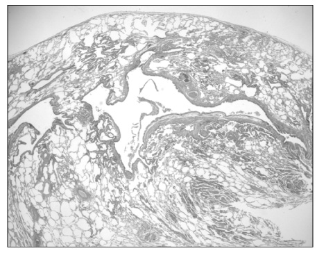
Figure 3 The resected lung shows various sized cystic lesion (H&E stain, ×10).

Figure 4 The lining cells are pseudostratified ciliated columnar cells to columnar and cuboidal cells like bronchiolar epithelial cells (H&E stain, ×100).

Figure 5 Focal mucinous epithelial lesion with mild atypism is revealed in the intervening parenchyma (H&E stain, ×200).
Cited by 1 articles
-
A Case of Atypical Adenomatous Hyperplasia of Larger Than 2 cm
Bo Mi Park, Min Ji Cho, Hyun Seok Lee, Dong Il Park, Myoung Rin Park, Ju Ock Kim, Jeong Eun Lee, Choong Sik Lee, Sung Soo Jung
Tuberc Respir Dis. 2013;74(6):280-285. doi: 10.4046/trd.2013.74.6.280.
Reference
-
1. Seo JS, Lee YC, Rhee YK. A case of congenital cystic adenomatoid malformation in adult patient. Korean J Med. 1993. 45:104–108.2. Stocker JT. Dali DH, Hammar SP, editors. Congenital and developmental diseases. Pulmonary pathology. 1994. 2nd ed. New York: Springer-verlag;155–190.3. Kim JI, Oh BJ, Song MH, Yun JP, Kim SH, Do KH, et al. Congenital cystic adenomatoid malformation of the lung presenting as hemoptysis in 49-year-old woman: a case report. Korean J Crit Care Med. 2004. 19:139–142.4. Weng S, Tsuchiya E, Satoh Y, Kitagawa T, Nakagawa K, Sugano H. Multiple atypical adenomatous hyperplasia of type II pneumonocytes and bronchiolo-alveolar carcinoma. Histopathology. 1990. 16:101–103.5. Brambilla E, Travis WD, Colby TV, Corrin B, Shimosato Y. The new World Health Organization classification of lung tumours. Eur Respir J. 2001. 18:1059–1068.6. Chapman AD, Kerr KM. The association between atypical adenomatous hyperplasia and primary lung cancer. Br J Cancer. 2000. 83:632–636.7. Stocker JT, Madewell JE, Drake RM. Congenital cystic adenomatoid malformation of the lung: classification and morphologic spectrum. Hum Pathol. 1977. 8:155–171.8. Shackelford GD, Siegel MJ. CT appearance of cystic adenomatoid malformations. J Comput Assist Tomogr. 1989. 13:612–616.9. Ioachimescu OC, Mehta AC. From cystic pulmonary airway malformation, to bronchioloalveolar carcinoma and adenocarcinoma of the lung. Eur Respir J. 2005. 26:1181–1187.10. Kerr KM. Pulmonary preinvasive neoplasia. J Clin Pathol. 2001. 54:257–271.11. Kawakami S, Sone S, Takashima S, Li F, Yang ZG, Maruyama Y, et al. Atypical adenomatous hyperplasia of the lung: correlation between high resolution CT findings and histopathologic features. Eur Radiol. 2001. 11:811–814.12. Yokose T, Doi M, Tanno K, Yamazaki K, Ochiai A. Atypical adenomatous hyperplasia of the lung in autopsy cases. Lung Cancer. 2001. 33:155–161.13. Yokose T, Ito Y, Ochiai A. High prevalence of atypical adenomatous hyperplasia of the lung in autopsy specimens from elderly patients with malignant neoplasms. Lung Cancer. 2000. 29:125–130.14. Nakahara R, Yokose T, Nagai K, Nishiwaki Y, Ochiai A. Atypical adenomatous hyperplasia of the lung: a clinicopathological study of 118 cases including cases with multiple atypical adenomatous hyperplasia. Thorax. 2001. 56:302–305.15. Takigawa N, Segawa Y, Nakata M, Saeki H, Mandai K, Kishino D, et al. Clinical investigation of atypical adenomatous hyperplasia of the lung. Lung Cancer. 1999. 25:115–121.
- Full Text Links
-
- Actions
-
Cited
- CITED
-
- Close
- Share
-
- Similar articles
-
- A case of congenital cystic adenomatoid malformation in adult patient
- CONGENITAL CYSTIC ADENOMATOID MALFORMATION TREATED WITH EMERGENCY OPERATION
- A case of multicystic dysplastic kidney and cystic adenomatoid malformation of the lung identified as incidental findings
- Congenital Cystic Adenomatoid Malformation Associated with Extralobar Pulmonary Sequestration: A case report
- Surgical treatment of congenital cystic adenomatoid malformation
