Spontaneous Intracerebral Hematoma from Transient Occult Carotid-cavernous Fistula: A Case Report
- Affiliations
-
- 1Department of Neurosurgery, College of Medicine, Wonkwang University, Iksan, Korea. kangsd@wonkwang.ac.kr
- 2Institute of Wonkwang Medical Science, Wonkwang University, Iksan, Korea.
- KMID: 1781748
- DOI: http://doi.org/10.3346/jkms.2005.20.1.166
Abstract
- After the spontaneous relief of initial symptoms by traumatic carotid-cavernous fistula (CCF), paradoxical worsening of patient's condition can be followed. We present a case of a 60-yr-old man whose audible bruit from a traumatic CCF had completely disappeared. A few days later, however, the patient had spontaneous intracerebral hematoma with cortical venous drainage. Complete obliteration of the fistula was achieved after embolization. When initial audible bruit in traumatic CCF disappears suddenly, cerebral angiography should be performed to differentiate venous hypertension by the hemodynamic changes of the cavernous sinus channels from spontaneous resolution of CCF.
MeSH Terms
Figure
-

Fig. 1 T2-weighted axial and sagittal magnetic resonance images show a dilated superior ophthalmic vein (arrows).
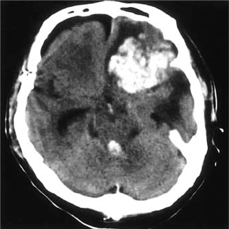
Fig. 2 Non contrast-enhanced computed tomography scan shows a large amount of hematoma in the left frontal base.

Fig. 3 Left internal carotid artery angiogram in lateral projection, showing direct CCF with drainage into sigmoid sinus (short arrow), superior ophthalmic vein (long arrow) and inferior ophthalmic vein (arrowhead), and dilated subfrontal pial veins (open arrow).
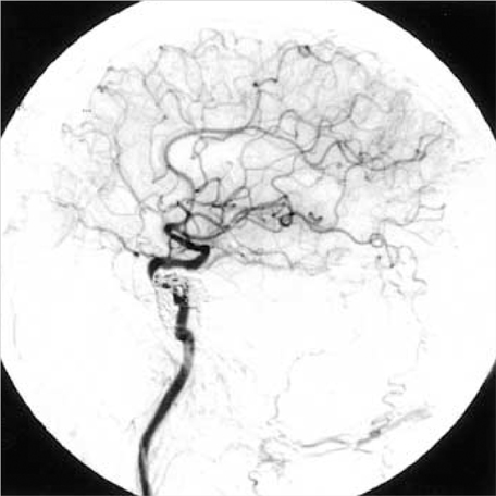
Fig. 4 Follow-up angiogram after the transarterial embolization shows a complete occlusion of CCF.
Cited by 1 articles
-
Cerebellar Hemorrhage due to a Direct Carotid–Cavernous Fistula after Surgery for Maxillary Cancer
Yoshinobu Kamio, Hisaya Hiramatsu, Mika Kamiya, Shuhei Yamashita, Hiroki Namba
J Korean Neurosurg Soc. 2017;60(1):89-93. doi: 10.3340/jkns.2015.1206.001.
Reference
-
1. d'Angelo VA, Monte V, Scialfa G, Fiumara E, Scotti G. Intracerebral venous hemorrhage in "high-risk" carotid cavernous fistula. Surg Neurol. 1988. 30:387–390.2. Halbach VV, Higashida RT, Hieshima GB, Reiche M, Norman D, Newton TH. Dural fistulas involving the cavernous sinus : results of treatment in 30 patients. Radiology. 1987. 163:437–442.3. Turner DM, Vangilder JC, Mojtahedi S, Pierson EW. Spontaneous intracerebral hematoma in carotid-cavernous fistula. Report of three cases. J Neurosurg. 1983. 59:680–686.4. Dandy WE. Carotid-cavernous aneurysms (pulsating exophthalmos). Zentralbl Neurochir. 1937. 2:77–113. 165–206.5. Locke CE. Intracranial arterio-venous aneurysm or pulsating exophthalmos. Ann Surg. 1924. 80:1–24.6. Martin JD, Mabson RF. Pulsating exophthalmos, review of all reported cases. JAMA. 1943. 121:330–335.7. van der Drift JH, Sparling CM, Berg D, Magnus O. Spontaneous occlusion of a carotid-cavernous shunt. Neurology. 1967. 17:187–193.
Article8. Isofort A. Spontangeilung einer trumatischen Carotissinus cavernousus-Fistel bei einem kind unter der Angiographue. Klin Mbl Augenhk. 1967. 150:821–823.9. Stenbens WE. Tindall G, Cooper PR, Barrow DL, editors. The pathogenesis of intracranial aneurysms. The practice of neurosurgery. 1996. Vol 2. Baltimore: Williams & Wilkins;1941–1952.10. Lin TK, Chang CN, Wai YY. Spontaneous intracerebral hematoma from occult carotid-cavernous fistula during pregnancy and puerperium. J Neurosurg. 1992. 76:714–717.
Article11. Muraine M, de Bokay E, Clavier E, Brasseur G. Paradoxical aggravation of carotid cavernous fistula. J Fr Ophthalmol. 1996. 19:51–54.
- Full Text Links
-
- Actions
-
Cited
- CITED
-
- Close
- Share
-
- Similar articles
-
- Traumatic Carotid-cavernous Fistula Bringing about Intracerebral Hemorrhage
- A Case of Spontaneous of Traumatic Carotid Cavernous Fistula After Carotid Angiography
- Delayed Spontaneous Thrombosis of Neglected Direct Carotid-Cavernous Fistula: A Case Report
- Delayed intracerebral hemorrhage from a traumatic carotid-cavernous fistula associated with an enucleated orbit
- Regional Cerebral Blood Flow Changes in Traumatic Carotid Cavernous Fistula During Trapping Procedure: Case Study, Preliminary Report
