Delayed intracerebral hemorrhage from a traumatic carotid-cavernous fistula associated with an enucleated orbit
- Affiliations
-
- 1Department of Neurosurgery, Neuropsychiatric Institute, University of Illinois at Chicago, Chicago, IL, USA
- KMID: 2520885
- DOI: http://doi.org/10.7461/jcen.2021.E2020.10.006
Abstract
- Spontaneous intracerebral hemorrhage (ICH) from a traumatic carotid-cavernous fistula (CCF) is a rare occurrence with few cases reported in the literature. Patients classically present shortly after the inciting trauma with symptoms of ocular venous hypertension. We report a case of an ICH due to delayed rupture of a venous aneurysm from a CCF in a patient with decades-old history of enucleation of the left globe secondary to trauma with no sentinel symptoms. Our patient represents a unique presentation of a rare pathology. This case highlights the need for ongoing surveillance in patients with a history of severe craniofacial trauma, as ICH from ruptured CCF(s) demands emergent treatment due to the potential for rapid neurological deterioration.
Keyword
Figure
-

Fig. 1. Axial CTA reveals cavernous sinus venous sacs and a dilated deep venous system (A). A venous network in the temporal lobe with connections to superficial cortical veins (B). Axial and coronal images show a periventricular venous varix arising from the thalamostriate vein (C, D). CTA, computed tomography angiogram.
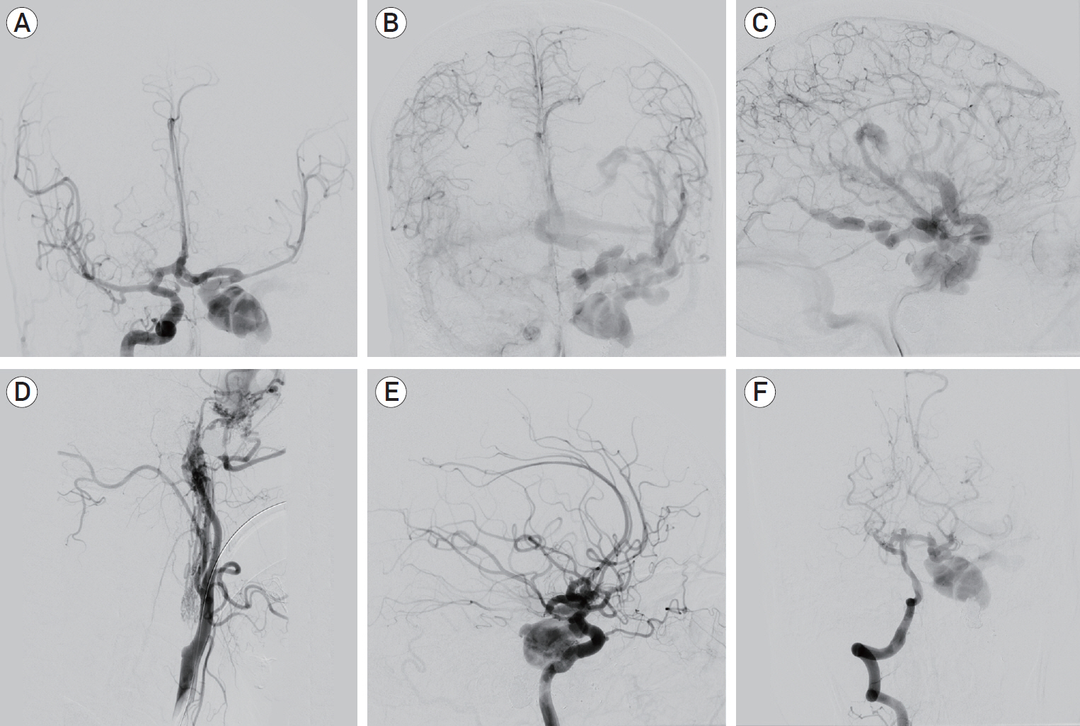
Fig. 2. AP DSA of the right ICA in the arterial phase shows rapid cross-flow through the anterior communicating artery complex into the CCF (A). There is evidence of venous hypertension as manifested by the dilation of both deep and superficial venous systems (B, C). Lateral DSA of the left CCA shows proximal ICA occlusion with supply of the CCF by way of ICA and ECA feeders (D). Oblique DSA of the right ICA and AP DSA of the right vertebral artery show flow-related aneurysms in the right A2 segment of the anterior cerebral artery and the basilar apex (E, F). AP, anteroposterior; DSA, digital subtraction angiography; ICA, internal carotid artery; CCF, carotid-cavernous fistula; CCA, common carotid artery; ECA, external carotid artery.
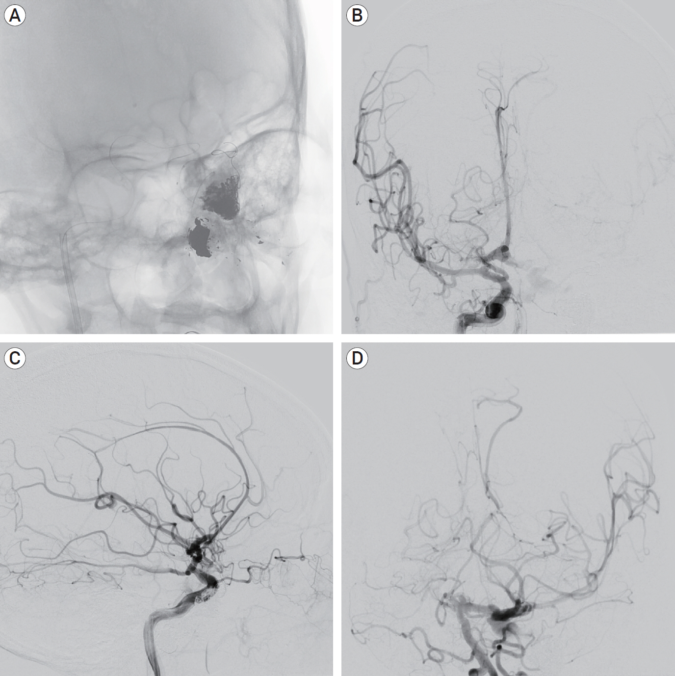
Fig. 3. Oblique unsubtracted view shows the combined Onyx-Coil mass (A). AP and lateral DSA of the right ICA after embolization shows near-complete occlusion of the CCF with no further evidence of retrograde venous reflux. The ACA territory fills from the right ICA bilaterally and there is faint filling of the left MCA territory from the right ICA injection (B, C). AP DSA of the left vertebral artery after embolization demonstrates filling of the left MCA territory by the posterior circulation via the posterior communicating artery (D). AP, anteroposterior; DSA, digital subtraction angiography; ICA, internal carotid artery; CCF, carotid-cavernous fistula; ACA, anterior cerebral artery; MCA, middle cerebral artery.
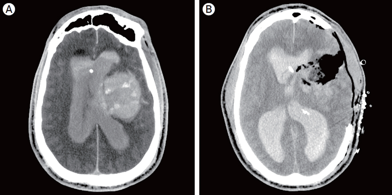
Fig. 4. Post-procedural axial computed tomography (CT) scan shows evidence of an expanding hematoma, IVH and midline shift (A). Post-operative axial CT scan after decompressive hemicraniectomy for hematoma evacuation (B). IVH, intraventricular hemorrhage.
Reference
-
1. Al-Mufti F, Amuluru K, El-Ghanem M, Changa A, Singh P, Gandhi C, et al. Spontaneous bilateral carotid-cavernous fistulas secondary to cavernous sinus thrombosis. Neurosurgery. 2017; Apr. 80(4):646–54.
Article2. Amuluru K, Al-Mufti F, Gandhi CD, Prestigiacomo CJ, Singh IP. Direct carotid-cavernous fistula: A complication of, and treatment with, flow diversion. Interv Neuroradiol. 2016; Oct. 22(5):569–76.
Article3. Aralasmak A, Karaali K, Cevikol C, Senol U, Sindel T, Toprak H, et al. Venous drainage patterns in carotid cavernous fistulas. ISRN Radiol. 2014; Jan. 2014:760267.
Article4. Barrow DL, Spector RH, Braun IF, Landman JA, Tindall SC, Tindall GT. Classification and treatment of spontaneous carotid-cavernous sinus fistulas. J Neurosurg. 1985; Feb. 62(2):248–56.
Article5. Chan FH, Shen CY, Liu JT, Li CS. Brainstem hemorrhage caused by direct carotid-cavernous fistula. A case report and literature review. Interv Neuroradiol. 2014; Jul-Aug. 20(4):487–94.6. Chang CM, Cheng CS. Late intracranial haemorrhage and subsequent carotid-cavernous sinus fistula after fracture of the facial bones. Br J Oral Maxillofac Surg. 2013; Dec. 51(8):e296–8.
Article7. Chavan RG, Kamble RB, Bonde V. Endovascular treatment in an unusual case of direct carotid cavernous fistula. Neuroradiol J. 2014; Apr. 27(2):207–12.
Article8. D’Angelo L, Paglia F, Caporlingua A, Sampirisi L, Guidetti G, Santoro A. Atypical manifestation of direct low-flow carotid-cavernous fistula: case report and review of the literature. World Neurosurg. 2019; May. 125:456–60.9. d’Angelo VA, Monte V, Scialfa G, Fiumara E, Scotti G. Intracerebral venous hemorrhage in “high-risk” carotid-cavernous fistula. Surg Neurol. 1988; Nov. 30(5):387–90.10. Halbach VV, Hieshima GB, Higashida RT, Reicher M. Carotid cavernous fistulae: indications for urgent treatment. AJR Am J Roentgenol. 1987; Sep. 149(3):587–93.
Article11. Hayashi K, Suyama K, Nagata I. Traumatic carotid cavernous fistula complicated with intracerebral hemorrhage: case report. Neurol Med Chir (Tokyo). 2011; 51(3):214–6.12. Hiramatsu K, Utsumi S, Kyoi K, Sakaki T, Tada T, Iwasaki S, et al. Intracerebral hemorrhage in carotid-cavernous fistula. Neuroradiology. 1991; 33(1):67–9.
Article13. Imrie A, Redmond K, Leggett D. Spontaneous direct carotid-cavernous sinus fistula secondary to a persistent primitive trigeminal artery treated by trans-venous coil embolisation. Interv Neuroradiol. 2018; Oct. 24(5):567–70.
Article14. Kamio Y, Hiramatsu H, Kamiya M, Yamashita S, Namba H. Cerebellar hemorrhage due to a direct carotid-cavernous fistula after surgery for maxillary cancer. J Korean Neurosurg Soc. 2017; Jan. 60(1):89–93.
Article15. Lang M, Habboub G, Mullin JP, Rasmussen PA. A brief history of carotid-cavernous fistula. J Neurosurg. 2017; Jun. 126(6):1995–2001.
Article16. Lee CJ, Choi SW, Kim SH, Youm JY. Traumatic carotid-cavernous fistula bringing about intracerebral hemorrhage. J Korean Neurosurgc Soc. 2005; Aug. 38(2):155–7.17. Leone G, Renieri L, Enriquez-Marulanda A, Dmytriw AA, Nappini S, Laiso A, et al. Carotid cavernous fistulas and dural arteriovenous fistulas of the cavernous sinus: Validation of a new classification according to venous drainage. World Neurosurg. 2019; Aug. 128:e621–31.
Article18. Lin TK, Chang CN, Wai YY. Spontaneous intracerebral hematoma from occult carotid-cavernous fistula during pregnancy and puerperium. Case report. J Neurosurg. 1992; Apr. 76(4):714–7.19. Moon KY, Kang SD. Spontaneous intracerebral hematoma from transient occult carotid-cavernous fistula: a case report. J Korean Med Sci. 2005; Feb. 20(1):166–8.20. Nagesh CP, Mohimen A, Kannath SK, Rajan JE. Primary intraventricular haemorrhage due to rupture of giant varix of the basal vein of Rosenthal in a patient with long-standing direct CCF: angiographic features and treatment considerations. BMJ Case Rep. 2017; Nov. 2017:bcr2017013396.
Article21. Tanaka A, Fukushima T, Tomonaga M. Intracerebral hematomas in cases of dural arteriovenous malformation and carotid-cavernous fistula. Surg Neurol. 1986; Jun. 25(6):557–62.
Article22. Thomas AJ, Chua M, Fusco M, Ogilvy C, Tubbs RS, Harrigan M, et al. Proposal of venous drainage – based classification system for carotid cavernous fistulae with validity assessment in a multicenter cohort. Neurosurgery. 2015; Sep. 77(3):380–5.23. Turner DM, Vangilder JC, Mojtahedi S, Pierson EW. Spontaneous intracerebral hematoma in carotid-cavernous fistula. Report of three cases. J Neurosurg. 1983; Oct. 59(4):680–6.24. Workman MJ, Dion JE, Tong FC, Cloft HJ. Treatment of Trapped CCF by Direct Puncture of the Cavernous Sinus by Infraocular Trans-SOF Approach. Case Report and Anatomical Basis. Interv Neuroradiol. 2002; Sep. 8(3):299–304.
- Full Text Links
-
- Actions
-
Cited
- CITED
-
- Close
- Share
-
- Similar articles
-
- Traumatic Carotid-cavernous Fistula Bringing about Intracerebral Hemorrhage
- Intracerebral Hemorrhage from a Traumatic Carotid Cavernous Fistula: Case Report
- Spontaneous Intracerebral Hematoma from Transient Occult Carotid-cavernous Fistula: A Case Report
- Bilateral Traumatic Carotid-Cavernous Fistula
- Regional Cerebral Blood Flow Changes in Traumatic Carotid Cavernous Fistula During Trapping Procedure: Case Study, Preliminary Report
