Epithelial Wound Healing after Cataract Surgery Comparing Two Different Topical Fluoroquinolones
- Affiliations
-
- 1Department of Ophthalmology, Hallym University College of Medicine, Chuncheon Sacred Heart Hospital, Chuncheon, Korea.
- 2Siloam Eye Hospital, Seoul, Korea.
- 3Corneal Dystrophy Research Institute, Department of Ophthalmology, Yonsei University College of Medicine, Seoul, Korea. eungkkim@yuhs.ac
- 4Department of Ophthalmology, Duke University, Durham, NC, USA.
- 5Severance Biomedical Science Institute, Yonsei University College of Medicine, Seoul, Korea.
- 6Brain Korea 21 Project for Medical Science, Yonsei University College of Medicine, Seoul, Korea.
- KMID: 1779906
- DOI: http://doi.org/10.3349/ymj.2014.55.1.197
Abstract
- PURPOSE
To compare the epithelial wound healing response of two preservative-free fluoroquinolones, moxifloxacin and levofloxacin, in patients who underwent cataract surgery.
MATERIALS AND METHODS
In this prospective, evaluator-masked, randomized clinical trial, 59 eyes of 50 patients who underwent cataract surgery were enrolled. Patients were randomized to receive moxifloxacin 0.5% (n=32 eyes) or levofloxacin 0.5% (n=27 eyes). All patients instilled moxifloxacin or levofloxain four times daily for 1 week prior to surgery and 2 weeks after surgery. The epithelial wound healing status in the corneal incision site was scanned with a raster scan mode of fourier-domain optical coherence tomography (FD-OCT). The number of eyes showing epithelial defect images and average number of corneal epithelial defect cuts per eye were compared between groups. All patients were evaluated on postoperative days 1, 2, 3, and 10.
RESULTS
On postoperative days 1, 2, and 3, the number of eyes showing epithelial defects in FD-OCT was not statistically different (all p>0.05). The average number of corneal epithelial defect cuts was also not statistically different between the two groups (all p>0.05). No eyes showed epithelial defects on postoperative day 10 in either group.
CONCLUSION
There were no differences on epithelial wound healing comparing these two different fluoroquinolones at the incision site of cataract surgery.
MeSH Terms
-
Aged
Aza Compounds/therapeutic use
Cataract Extraction
Cornea/drug effects/*surgery
Female
Fluoroquinolones/*therapeutic use
Humans
Levofloxacin/therapeutic use
Male
Middle Aged
Quinolines/therapeutic use
Tomography, Optical Coherence
Wound Healing/*drug effects
Aza Compounds
Fluoroquinolones
Levofloxacin
Quinolines
Figure
-
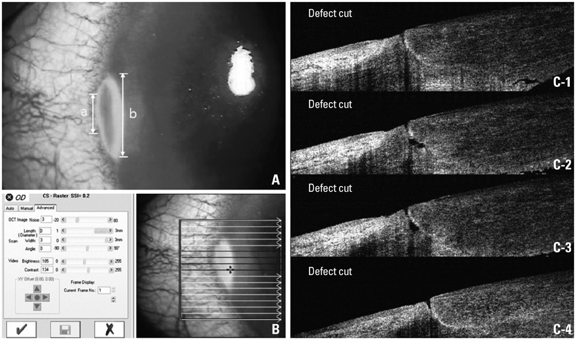
Fig. 1 (A) A slit lamp photograph of the corneal incision after fluorescence dye staining. The margin of corneal defect is obscure (a or b) and can be interpreted differently according to varying strengths of illumination. (B) A capture window of raster scan mode in fourier-domain optical coherence tomography (FD-OCT). By selecting a 3×3 mm2 sized capture window, 17 cuts of cross-sectional images (white and red arrows) could be acquired at the same gap. (C) On cross-sectional images of red arrows in Fig. 1B, the defect cuts (C-1 to 4) can be counted. The actual margin of corneal defect detected based on the FD-OCT images was determined to be "a" not "b" in Fig. 1A.

Fig. 2 (A) Representative images showing the defect and non-defect image cuts in images of the fourier-domain optical coherence tomography. (A and B) An imaginary line (red dotted line) was drawn connecting Bowman layer on both ends of the sectional plane. (A-1 and A-2) Cases showing a lack of epithelium on the imaginary line and a discontinuation of this line were interpreted as a defect cut. The black hollow spaces without epithelial growth were noted (white arrows). The layer of powerfully bright light is considered to be a reflection of the tear fluid (white hollow arrowheads). (B-1 and B-2) Cases showing epithelial growth both above this line and without interruptions were interpreted as a non-defect image cut. The spaces in the incision site were occupied with epithelial growth which showing similar dark gray color with the epithelial layer (black arrows).
Reference
-
1. Hooper DC, Wolfson JS. Mode of action of the quinolone antimicrobial agents. Rev Infect Dis. 1988; 10:Suppl 1. S14–S21.
Article2. Mather R, Karenchak LM, Romanowski EG, Kowalski RP. Fourth generation fluoroquinolones: new weapons in the arsenal of ophthalmic antibiotics. Am J Ophthalmol. 2002; 133:463–466.
Article3. Vargas LG, Vroman DT, Solomon KD, Holzer MP, Escobar-Gomez M, Schmidbauer JM, et al. Epithelial downgrowth after clear cornea phacoemulsification: report of two cases and review of the literature. Ophthalmology. 2002; 109:2331–2335.
Article4. Lee BL, Gaton DD, Weinreb RN. Epithelial downgrowth following phacoemulsification through a clear cornea. Arch Ophthalmol. 1999; 117:283.
Article5. Pollock GA, McKelvie PA, McCarty DJ, White JF, Mallari PL, Taylor HR. In vivo effects of fluoroquinolones on rabbit corneas. Clin Experiment Ophthalmol. 2003; 31:517–521.
Article6. Patel GM, Chuang AZ, Kiang E, Ramesh N, Mitra S, Yee RW. Epithelial healing rates with topical ciprofloxacin, ofloxacin, and ofloxacin with artificial tears after photorefractive keratectomy. J Cataract Refract Surg. 2000; 26:690–694.
Article7. Moreira LB, Lee RF, de Oliveira C, LaBree L, McDonnell PJ. Effect of topical fluoroquinolones on corneal re-epithelialization after excimer laser keratectomy. J Cataract Refract Surg. 1997; 23:845–848.
Article8. Moshirfar M, Chew J, Werner L, Meyer JJ, Hunter B, Stevens S, et al. Comparison of the effects of fourth-generation fluoroquinolones on corneal re-epithelialization in rabbit eyes. Graefes Arch Clin Exp Ophthalmol. 2008; 246:1455–1461.
Article9. Kowalski RP, Kowalski BR, Romanowski EG, Mah FS, Thompson PP, Gordon YJ. The in vitro impact of moxifloxacin and gatifloxacin concentration (0.5% vs 0.3%) and the addition of benzalkonium chloride on antibacterial efficacy. Am J Ophthalmol. 2006; 142:730–735.
Article10. Tsai TH, Chen WL, Hu FR. Comparison of fluoroquinolones: cytotoxicity on human corneal epithelial cells. Eye (Lond). 2010; 24:909–917.
Article11. Sosa AB, Epstein SP, Asbell PA. Evaluation of toxicity of commercial ophthalmic fluoroquinolone antibiotics as assessed on immortalized corneal and conjunctival epithelial cells. Cornea. 2008; 27:930–934.
Article12. Kim SY, Lim JA, Choi JS, Choi EC, Joo CK. Comparison of antibiotic effect and corneal epithelial toxicity of levofloxacin and moxifloxacin in vitro. Cornea. 2007; 26:720–725.
Article13. Kovoor TA, Kim AS, McCulley JP, Cavanagh HD, Jester JV, Bugde AC, et al. Evaluation of the corneal effects of topical ophthalmic fluoroquinolones using in vivo confocal microscopy. Eye Contact Lens. 2004; 30:90–94.
Article14. Watanabe R, Nakazawa T, Yokokura S, Kubota A, Kubota H, Nishida K. Fluoroquinolone antibacterial eye drops: effects on normal human corneal epithelium, stroma, and endothelium. Clin Ophthalmol. 2010; 4:1181–1187.
Article
- Full Text Links
-
- Actions
-
Cited
- CITED
-
- Close
- Share
-
- Similar articles
-
- Effects of 0.1% Dexamethasone on Experimental corneal Epithelial Healing Following Alkali Wounds
- Effect of Epidermal Growth Factor on the Wound Healing after Excimer Laser Photokeratectomy in Rabbits
- Corneal Epithelial Wound Healing After Treatment with Gatifloxacin with or Without Benzalkonium Chloride in Rabbits
- The Effect of Topical Prostaglandin Synthesis Inhibitor and Therapeutic Contact Lens on the Pain and Corneal Reepithelialization after Excimer Laser PRK
- Homologous fibronectin enhances healing of excised wounds in rats
