Sonographically-Guided 14-Gauge Core Needle Biopsy for Papillary Lesions of the Breast
- Affiliations
-
- 1Department of Radiology, Seoul National University College of Medicine, Institute of Radiation Medicine, Seoul National University Medical Research Center, Seoul, Korea. moonwk@radcom.snu.ac.kr
- KMID: 1779435
- DOI: http://doi.org/10.3348/kjr.2007.8.3.206
Abstract
OBJECTIVE
We wanted to assess the need for surgical excising papillary lesions of the breast that were diagnosed upon sonographically guided 14-gauge core needle biopsy. MATERIALS AND METHODS: Sixty-nine women (age range: 25-74 years, mean age: 51.7 years) with 69 papillary lesions (4.9%) were diagnosed and followed after performing sonographically guided 14-gauge core needle biopsies. Surgical excision was performed for 44 (64%) of 69 papillary lesions, and 25 lesions were followed with imaging studies (range: 6-46 months, mean: 17.9 months). The histologic findings upon core biopsy were compared with the surgical, imaging and follow-up findings. RESULTS: Core needle biopsies of 69 lesions yielded tissue that was classified as benign for 43 lesions, atypical for 18 lesions and malignant for eight lesions. Of the 43 lesions that yielded benign papilloma upon core needle biopsy, one had intraductal papillary carcinoma found upon surgery. An immediate surgical biopsy was recommended for this lesion because of the imaging-histologic discordance. No additional carcinoma was found during the imaging follow-up. Surgical excision was performed for 17 atypical papillary lesions, and this revealed intraductal (n = 6) or invasive (n = 2) papillary carcinoma in 8 (47%) lesions. Of the seven intraductal papillary carcinomas, surgery revealed invasive papillary carcinoma in one (14%). CONCLUSION: Our results suggest that papillary lesions of the breast that are diagnosed as benign upon sonographically guided 14-gauge core needle biopsy can be followed when the results are concordant with the imaging findings.
Keyword
MeSH Terms
Figure
-
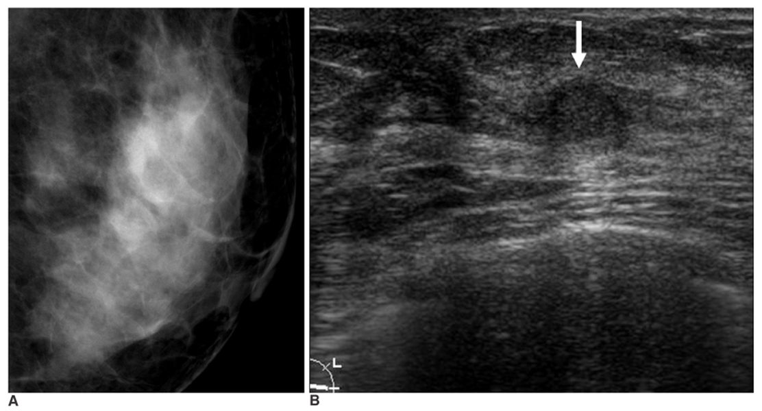
Fig. 1 An asymptomatic 52-year-old woman with benign papilloma upon core needle biopsy. A. Collimated photograph of the mediolateral oblique mammogram reveals no focal abnormality in the left subareolar area. B. Sonogram reveals a 7 mm oval mass (arrow) in the left subareolar area. This finding was thought to be concordant with the benign histology of the core biopsy. A subsequent surgical rebiopsy due to the patient's concern also revealed benign papilloma.
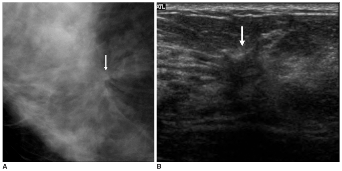
Fig. 2 An asymptomatic 34-year-old woman with benign papilloma upon core needle biopsy. A. Collimated photograph of the craniocaudal mammogram reveals a 2 cm area of architectural distortion with central density (arrow) in the right subareolar area. B. Sonogram reveals a 1.6 cm irregular hypoechoic mass (arrow) with a spiculated margin and ductal extension. A subsequent surgical rebiopy was recommended due to the imaging-histologic discordance, and it revealed intraductal papillary carcinoma.
Cited by 1 articles
-
Is US-guided 14-gauge Core Needle Biopsy Valid for Papillary Neoplasm of the Breast?
Na Young Jung, Jaehee Lee, A Won Lee, Byung Joo Song, Sang Seol Jung
J Breast Cancer. 2008;11(1):30-35. doi: 10.4048/jbc.2008.11.1.30.
Reference
-
1. Rosen PP. Rosen PP, editor. Benign papillary tumors. Rosen's breast pathology. 1997. Philadelphia: Lippincott-Raven;67–104.2. Rosen PP. Rosen PP, editor. Papillary carcinoma. Rosen's breast pathology. 1997. Philadelphia: Lippincott-Raven;335–354.3. Gadd MA. Harris JR, Lippman ME, Morrow M, Hellman S, editors. Papillary lesions. Diseases of the breast. 1996. Philadelphia: Lippincott-Raven;42–45.4. Haagensen CD. Haagensen CD, editor. Solitary intraductal papilloma. Diseases of the breast. 1986. Philadelphia: Saunders;136–175.5. Ellis IO, Elston CW, Pinder SE. Elston CW, Ellis IO, editors. Papillary lesions. Systemic pathology: the breast. 1998. 3rd ed. Edinburgh: Churchill Livingstone;133–146.6. Tavassoli FA. Tavassoli FA, editor. Papillary lesions. Pathology of the breast. 1992. New York: Elsevier, Norwalk; Conn;193–227.7. Liberman L, Bracero N, Vuolo MA, Dershaw DD, Morris EA, Abramson AF, et al. Percutaneous large-core biopsy of papillary breast lesions. AJR Am J Roentgenol. 1999. 172:331–337.8. Rosen EL, Bentley RC, Baker JA, Soo MS. Imaging-guided core needle biopsy of papillary lesions of the breast. AJR Am J Roentgenol. 2002. 179:1185–1192.9. Liberman L, Feng TL, Dershaw DD, Morris EA, Abramson AF. US-guided core breast biopsy: use and cost-effectiveness. Radiology. 1998. 208:717–723.10. Parker SH, Jobe WE, Dennis MA, Stavros AT, Johnson KK, Yakes WF, et al. US-guided automated large-core breast biopsy. Radiology. 1993. 187:507–511.11. American College of Radiology. Breast imaging reporting and data system (BI-RADSTM) - Mammography. 2003. Reston, Va: American College of Radiology.12. American College of Radiology. Breast imaging reporting and data system (BI-RADSTM) - Ultrasound. 2003. Reston, Va: American College of Radiology.13. Gisvold JJ, Goellner JR, Grant CS, Donohue JH, Skyes MW, Karsell PR, et al. Breast biopsy: a comparative study of stereotaxically guided core and excisional techniques. AJR Am J Roentgenol. 1994. 162:815–820.14. Parker SH, Burbank F, Jackman RJ, Aucreman CJ, Cardenosa G, Cink TM, et al. Percutaneous large-core needle biopsy: a multi-institutional study. Radiology. 1994. 193:359–364.15. Cho N, Moon WK, Cha JH, Kim SM, Kim SJ, Lee SH, et al. Sonographically guided core biopsy of the breast: comparison of 14-gauge automated gun and 11-gauge directional vacuum-assisted biopsy methods. Korean J Radiol. 2005. 6:102–109.16. Burbank F. Stereotactic breast biopsy of atypical ductal hyperplasia and ductal carcinoma in situ lesions: improved accuracy with directional, vacuum-assisted biopsy. Radiology. 1997. 202:843–847.17. Jackman RJ, Nowels KW, Rodriguez-Soto J, Marzoni FA Jr, Finkelstein SI, Shepard MJ. Stereotactic, automated, large-core needle biopsy of nonpalpable breast lesions: false-negative and histologic underestimation rates after long-term follow-up. Radiology. 1999. 210:799–805.18. Mercado CL, Hamele-Bena D, Singer C, Koenigsberg T, Pile-Spellman H, Higgins H, et al. Papillary lesions of the breast: evaluation with stereotactic directional vacuum-assisted biopsy. Radiology. 2001. 221:650–655.19. Buchberger W, DeKoekkoek-Doll P, Springer P, Obrist P, Dunser M. Incidental findings on sonography of the breast: clinical significance and diagnostic workup. AJR Am J Roentgenol. 1999. 173:921–927.20. Kolb TM, Lichy J, Newhouse JH. Occult cancer in women with dense breasts: detection with screening US-diagnostic yield and tumor characteristics. Radiology. 1998. 207:191–199.21. Moon WK, Noh DY, Im JG. Multifocal, multicentric, and contralateral breast cancers: bilateral whole-breast US in the preoperative evaluation of patients. Radiology. 2002. 224:569–576.22. Mercado CL, Hamele-Bena D, Oken SM, Singer CI, Cangiarella J. Papillary lesions of the breast at percutaneous core-needle biopsy. Radiology. 2006. 238:801–808.23. Liberman L, Tornos C, Huzjan R, Bartella L, Morris EA, Dershaw DD. Is surgical excision warranted after benign, concordant diagnosis of papilloma at percutaneous breast biopsy? AJR Am J Roentgenol. 2006. 186:1328–1334.
- Full Text Links
-
- Actions
-
Cited
- CITED
-
- Close
- Share
-
- Similar articles
-
- Usefulness of Ultrasound-Guided Automated Core Biopsy of Nonpalpable Breast Lesions
- Clinicopathologic Features of the Papillary Breast Lesions Diagnosed on Ultrasonography-guided Core Needle Biopsy
- Track Seeding in a Breast Cancer Patient after a 14-Gauge Core Needle Biopsy: A Case Report
- Phyllodes Tumors and Fibroepithelial Lesions with Cellular Stroma of the Breast and Diagnosed by Sonographically Guided Core Needle Biopsy: A Comparison Between the Results of Excision Biopsy and the Sonographic Findings
- Is US-guided 14-gauge Core Needle Biopsy Valid for Papillary Neoplasm of the Breast?
