J Korean Med Sci.
2009 Apr;24(2):346-349. 10.3346/jkms.2009.24.2.346.
Perivascular Epithelioid Cell Tumor (PEComa) of Abdominal Cavity from Falciform Ligament: A Case Report
- Affiliations
-
- 1Department of Internal Medicine, Pusan National University College of Medicine, Busan, Korea.
- 2Department of Radiology, Pusan National University College of Medicine, Busan, Korea. kto0440@yahoo.co.kr
- 3Department of General Surgery, Pusan National University College of Medicine, Busan, Korea.
- KMID: 1779144
- DOI: http://doi.org/10.3346/jkms.2009.24.2.346
Abstract
- We present a case of perivascular epithelioid cell tumors (PEComas) in the abdominal cavity at the falciform ligament. A 30-yr-old Korean man visited to hospital for the evaluation of a growing, palpable abdominal mass. He had felt the mass growing over 6 months. There was no family or personal history of tuberous sclerosis. The resected specimen showed a mass of 8.0x7.0x5.5 cm in size. Histological examination showed sheets of spindle-to-epithelioid cells with clear-to-eosinophilic cytoplasm. Immunohistochemically, tumor cells were positive for HMB-4 (gp100) and smooth muscle actin. They were also positive for the S-100, which is a marker of neurogenic and melanocytic tumors. Patient was treated with radical resection of tumor without any adjuvant therapy. He is well and on follow-up visits without tumor recurrence.
Keyword
MeSH Terms
Figure
-
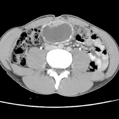
Fig. 1 Abdominal computed tomography. Well enhancing mural nodule and wall calcification were noted.
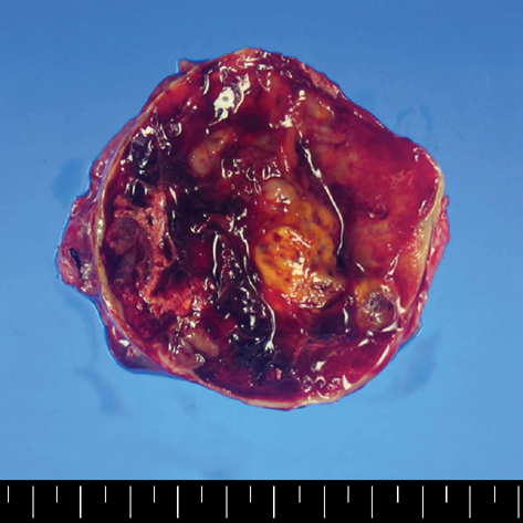
Fig. 2 Gross finding. On section it showed a uniloculated cystic mass with solid portion. The solid portion was yellow brown color and it revealed foci of hemorrhage and necrotic change.
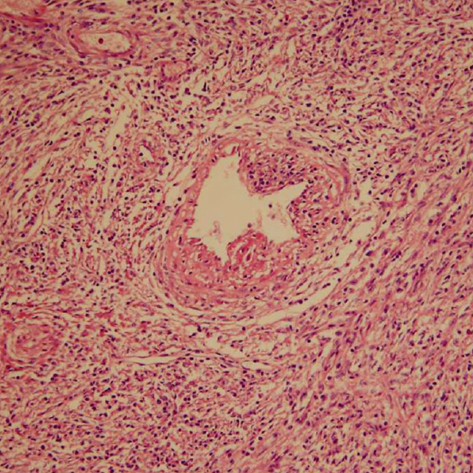
Fig. 3 Histologic findings (H&E stain, ×100). The lesion was made of sheets of spindle-to-epithelioid cells on perivascular space. The spindle cells had clear-to-eosinophilic cytoplasm.
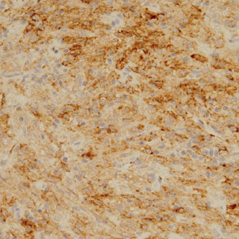
Fig. 4 Immunohistochemical stain. Tumor cells were positive for HMB-45.
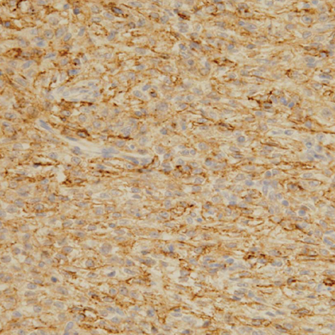
Fig. 5 Immunohistochemical stain. Tumor cells were positive for SMA.
Reference
-
1. Folpe AL, Mentzel T, Lehr HA, Fisher C, Balzer BL, Weiss SW. Perivascular epithelioid cell neoplasms of soft tissue and gynecologic origin: a clinicopathologic study of 26 cases and review of the literature. Am J Surg Pathol. 2005. 29:1558–1575.2. Pea M, Bonetti F, Zamboni G, Martignoni G, Riva M, Colombari R, Mombello A, Bonzanini M, Scarpa A, Ghimenton C. Melanocyte-marker-HMB-45 is regularly expressed in angiomyolipoma of the kidney. Pathology. 1991. 23:185–188.
Article3. Pea M, Bonetti F, Zamboni G, Martignoni G, Fiore-Donati L, Doglioni C. Clear cell tumor and angiomyolipoma. Am J Surg Pathol. 1991. 15:199–202.
Article4. Weeks DA, Malott RL, Arnesen M, Zuppan C, Aitken D, Mierau G. Hepatic angiomyolipoma with striated granules and positivity with melanoma--specific antibody (HMB-45): a report of two cases. Ultrastruct Pathol. 1991. 15:563–571.
Article5. Gaffey MJ, Mills SE, Zarbo RJ, Weiss LM, Gown AM. Clear cell tumor of the lung. Immunohistochemical and ultrastructural evidence of melanogenesis. Am J Surg Pathol. 1991. 15:644–653.
Article6. Gal AA, Koss MN, Hochholzer L, Chejfec G. An immunohistochemical study of benign clear cell ('sugar') tumor of the lung. Arch Pathol Lab Med. 1991. 115:1034–1038.7. Bonetti F, Pea M, Martignoni G, Zamboni G, Iuzzolino P. Cellular heterogeneity in lymphangiomyomatosis of the lung. Hum Pathol. 1991. 22:727–728.
Article8. Bonetti F, Pea M, Martignoni G, Zamboni G. PEC and sugar. Am J Surg Pathol. 1992. 16:307–308.
Article9. Martignoni G, Pea M, Reghellin D, Zamboni G, Bonetti F. Perivascular epithelioid cell tumor (PEComa) in the genitourinary tract. Adv Anat Pathol. 2007. 14:36–41.
Article10. Al-Saleem T, Wessner LL, Scheithauer BW, Patterson K, Roach ES, Dreyer SJ, Fujikawa K, Bjornsson J, Bernstein J, Henske EP. Malignant tumors of the kidney, brain, and soft tissues in children and young adults with the tuberous sclerosis complex. Cancer. 1998. 83:2208–2216.
Article11. Harris GC, McCulloch TA, Perks G, Fisher C. Malignant perivascular epithelioid cell tumour ("PEComa") of soft tissue: a unique case. Am J Surg Pathol. 2004. 28:1655–1658.12. Fink D, Marsden DE, Edwards L, Camaris C, Hacker NF. Malignant perivascular epithelioid cell tumor (PEComa) arising in the broad ligament. Int J Gynecol Cancer. 2004. 14:1036–1039.
Article13. Lehman NL. Malignant PEComa of the skull base. Am J Surg Pathol. 2004. 28:1230–1232.
Article14. Yanai H, Matsuura H, Sonobe H, Shiozaki S, Kawabata K. Perivascular epithelioid cell tumor of the jejunum. Pathol Res Pract. 2003. 199:47–50.
Article15. Pan CC, Yang AH, Chiang H. Malignant perivascular epithelioid cell tumor involving the prostate. Arch Pathol Lab Med. 2003. 127:E96–E98.
Article16. Evert M, Wardelmann E, Nestler G, Schulz HU, Roessner A, Rocken C. Abdominopelvic perivascular epithelioid cell sarcoma (malignant PEComa) mimicking gastrointestinal stromal tumour of the rectum. Histopathology. 2005. 46:115–117.
Article17. Folpe AL, Goodman ZD, Ishak KG, Paulino AF, Taboada EM, Meehan SA, Weiss SW. Clear cell myomelanocytic tumor of the falciform ligament/ligamentum teres: a novel member of the perivascular epithelioid clear cell family of tumors with a predilection for children and young adults. Am J Surg Pathol. 2000. 24:1239–1246.18. Birkhaeuser F, Ackermann C, Flueckiger T, Guenin MO, Kern B, Tondelli P, Peterli R. First description of a PEComa (perivascular epithelioid cell tumor) of the colon: report of a case and review of the literature. Dis Colon Rectum. 2004. 47:1734–1737.
Article
- Full Text Links
-
- Actions
-
Cited
- CITED
-
- Close
- Share
-
- Similar articles
-
- Primary Perivascular Epithelioid Cell Tumor (PEComa) of the Liver: A Case Report and Review of the Literature
- Malignant Perivascular Epithelioid Cell Tumor (PEComa) Arising in the Omentum with Metastatic Peritoneal Seeding: A Case Report
- Perivascular epithelioid cell tumor (PEComa) of the ascending colon: the implication of IFN-alpha2b treatment
- A case of perivascular epithelioid cell tumor of the uterus
- Pigmented Perivascular Epithelioid Cell Tumor (PEComa) of the Kidney: A Case Report and Review of the Literature
