J Korean Med Sci.
2005 Jun;20(3):483-488. 10.3346/jkms.2005.20.3.483.
Development of Korean Standard Brain Templates
- Affiliations
-
- 1Korean Consortium for Brain Mapping, Department of Nuclear Medicine, Seoul National University College of Medicine, Seoul, Korea. dsl@plaza.snu.ac.kr
- 2Department of Neuropsychiatry, Seoul National University College of Medicine, Seoul, Korea.
- 3Department of Family Practice, Seoul National University College of Medicine, Seoul, Korea.
- 4Department of Radiology, Seoul National University College of Medicine, Seoul, Korea.
- 5Department of Nuclear Medicine, National Cancer Center, Goyang, Korea.
- 6Department of Biomedical Engineering, Hanyang University, Seoul, Korea.
- 7Department of Neuropsychiatry, Yonsei University College of Medicine, Gwangju, Korea.
- KMID: 1778511
- DOI: http://doi.org/10.3346/jkms.2005.20.3.483
Abstract
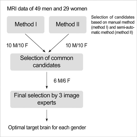
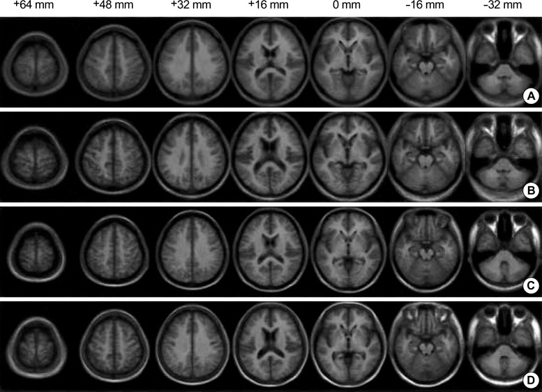
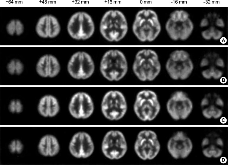
- We developed age, gender and ethnic specific brain templates based on MR and Positron-Emission Tomography (PET) images of Korean normal volunteers. Seventy-eight normal right-handed volunteers (M/F=49/29) underwent 3D T1-weighted SPGR MR and F-18-FDG PET scans. For the generation of standard templates, an optimal target brain that has the average global hemispheric shape was selected for each gender. MR images were then spatially normalized by linear transformation to the target brains, and normalization parameters were reapplied to PET images. Subjects were subdivided into 2 groups for each gender: the young/midlife (<55 yr) and the elderly groups. Young and elderly MRI/PET templates were composed by averaging the spatially normalized images. Korean templates showed different shapes and sizes (mean length, width, and height of the brains were 16.5, 14.3 and 12.1 cm for man, and 15.6, 13.5 and 11.4 cm for woman) from the template based on Caucasian (18.3, 14.2, and 13.3 cm). MRI and PET templates developed in this study will provide the framework for more accurate stereotactic standardization and anatomical localization.
Keyword
MeSH Terms
Figure
Reference
-
1. Kasai K, Iwanami A, Yamasue H, Kuroki N, Nakagome K, Fukuda M. Neuroanatomy and neurophysiology in schizophrenia. Neurosci Res. 2002. 43:93–110.
Article2. Demetriades AK. Functional neuroimaging in Alzheimer's type dementia. J Neurol Sci. 2002. 203-204:247–251.
Article3. Theodore WH, Gaillard WD. Neuroimaging and the progression of epilepsy. Prog Brain Res. 2002. 135:305–313.
Article4. Goldstein RZ, Volkow ND. Drug addiction and its underlying neurobiological basis: neuroimaging evidence for the involvement of the frontal cortex. Am J Psychiatry. 2002. 159:1642–1652.
Article5. Munte TF, Altenmuller E, Jancke L. The musician's brain as a model of neuroplasticity. Nat Rev Neurosci. 2002. 3:473–478.
Article6. Lee DS, Lee JS, Oh SH, Kim SK, Kim JW, Chung JK, Lee MC, Kim CS. Cross-modal plasticity and cochlear implants. Nature. 2001. 409:149–150.
Article7. Giraud AL, Truy E, Frackowiak R. Imaging plasticity in cochlear implant patients. Audiol Neurootol. 2001. 6:381–393.
Article8. Evans AC, Collins DL, Mills SR, Brown ED, Kelly RL, Peters TM. 3D statistical neuroanatomical models from 305 MRI volumes. Proc IEEE Nucl Science Symp Medl Imaging Conf. 1993. In : Nuclear Science Symposium and Medical Imaging Conference; 10/31/1993 - 11/06/1993; San Francisco, CA, USA. 1813–1817.
Article9. Ashburner J, Friston KJ. Nonlinear spatial normalization using basis functions. Hum Brain Mapp. 1999. 7:254–266.
Article10. Lancaster JL, Fox PT, Downs H, Nickerson DS, Hander TA, El Mallah M, Kochunov PV, Zamarripa F. Global spatial normalization of human brain using convex hulls. J Nucl Med. 1999. 40:942–955.11. Toga AW, Thompson PM. Maps of the brain. Anat Rec. 2001. 265:37–53.
Article12. Mazziotta J, Toga A, Evans A, Fox P, Lancaster J, Zilles K, Woods R, Paus T, Simpson G, Pike B, Holmes C, Collins L, Thompson P, MacDonald D, Iacoboni M, Schormann T, Amunts K, Palomero-Gallagher N, Geyer S, Parsons L, Narr K, Kabani N, Le Goualher G, Boomsma D, Camon T, Kawashima R, Mazoyer B. A probabilistic atlas and reference system for the human brain: International Consortium for Brain Mapping (ICBM). Philos Trans R Soc Lond B Biol Sci. 2001. 356:1293–1322.
Article13. Talairach J, Tournoux P. Co-planar stereotaxic atlas of the human brain: 3-dimensional proportional system-an approach to cerebral imaging. 1988. New York: Thieme Medical Publishers.14. Fox PT, Perlmutter JS, Raichle ME. A stereotactic method of anatomical localization for positron emission tomography. J Comput Assist Tomogr. 1985. 9:141–153.
Article15. Friston KJ, Holmes AP, Worsley KJ, Poline JP, Frith CD, Frackowiak RS. Statistical parametric maps in functional imaging: a general linear approach. Hum Brain Mapp. 1995. 2:189–210.
Article16. Tzourio-Mazoyer N, Landeau B, Papathanassiou D, Crivello F, Etard O, Delcroix N, Mazoyer B, Joliot M. Automated anatomical labeling of activations in SPM using a macroscopic anatomical parcellation of the MNI MRI single-subject brain. Neuroimage. 2002. 15:273–289.
Article17. Yoon U, Lee JM, Kim JJ, Lee SM, Kim IY, Kwon JS, Kim SI. Modified magnetic resonance image based parcellation method for cerebral cortex using successive fuzzy clustering and boundary detection. Ann Biomed Eng. 2003. 31:441–447.
Article18. Guimond A, Meunier J, Thirion JP. Average brain models: a convergence study. Comput Vision Imag Understand. 2000. 77:192–201.
Article19. Kochunov P, Lancaster J, Thompson P, Toga AW, Brewer P, Hardies J, Fox P. An optimized individual target brain in the Talairach coordinate system. Neuroimage. 2002. 17:922–927.
Article
- Full Text Links
- Actions
-
Cited
- CITED
-
- Close
- Share
- Similar articles
-
- Constructing the KOR152 Korean Young Adult Brain Atlas Utilizing the State-of-the-Art Method for the Age-Specific Population
- Development of Korean Standard Brain Templates According to Gender and Age
- Quantification of Brain Images Using Korean Standard Templates and Structural and Cytoarchitectonic Probabilistic Maps
- The application of template constructed by cephalometric roentgenograms in Hellman dental age IV A with normal occlusion
- Development of a Korean Standard Structural Brain Template in Cognitive Normals and Patients with Mild Cognitive Impairment and Alzheimer's Disease


